


![]()
News
Last updated 6 May 2026
Wednesday 6 May 2026
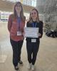
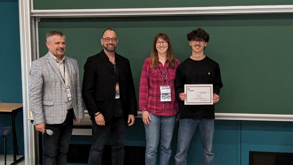
During the Journées RITS held at the University of Strasbourg (April 20–22, 2026), researchers from the LabTAU showcased a strong and diverse set of contributions highlighting their work in therapeutic ultrasound and biomedical imaging.
Special congratulations to Marine SIMONNEAU for receiving the Best Oral Presentation Award, recognizing the quality and impact of her work!
Well done to all contributors for representing LabTAU and advancing the field of biomedical acoustics.

Thursday 12 February 2026
Author: Mouna GUIZANI
Time: 14H00
Language: French
Place: Conference Room at LabTAU
Abstract: This thesis investigates the flow generated by focused ultrasound within a fluid confined in a circular tube, in a configuration compatible with a centimetric human blood vessel. Experimental and numerical investigations are conducted on simplified model of a rigid, straight circular tube, both in the case of a closed conduit and in the case of an imposed steady flow. Experimentally, a setup based on a high-intensity focused ultrasound (HIFU) therapy transducer was implemented to generate a focused ultrasound beam parallel to the tube axis. Velocity field measurements were performed in water using particle image velocimetry (PIV). In parallel, numerical simulations based on the experimental HIFU field and modeling its fluidic effect as a volumic force field were carried out using OpenFOAM software, first for a Newtonian fluid and then for a non-Newtonian fluid modeled by the Bird-Carreau law.
The vortex structure generated by focused ultrasound is characterized by a dense core of streaming flow aligned with the direction of the ultrasonic wave, accompanied by a low-velocity reverse flow. While the position of the ultrasound focus within the tube is not critical for generating mixing, it significantly influences the wall shear stress, which increases as the focus approaches the tube wall. The comparison between Newtonian and Bird-Carreau fluid revealed that at high acoustic intensities, the Bird-Carreau fluid behaves similarly to the Newtonian fluid, maintaining effective mixing and wall shear stress. Further investigation, focusing on the case where ultrasound cavitation occurs under the influence of the HIFU field, enabled the estimation of an effective ultrasound attenuation coefficient characterizing the cavitating medium.
Titre : Contrôle par ultrasons focalisés d’un écoulement en conduite en vue de traitements vasculaires.
Résumé : La présente thèse étudie l'écoulement induit par des ultrasons focalisés au sein d’un fluide confiné en conduite, dans une configuration compatible avec un vaisseau sanguin humain de calibre centimétrique. Les investigations, expérimentales et numériques, sont menées sur le modèle simplifié d’un tube circulaire droit indéformable, à la fois dans le cas d’un conduit fermé et celui d’un débit permanent imposé. Sur le plan expérimental, un dispositif basé sur un transducteur de thérapie par ultrasons focalisés de haute intensité (HIFU) a été mis en œuvre afin de générer un faisceau ultrasonore focalisé parallèle à l’axe du tube. Des mesures de champs de vitesse ont été réalisées dans l’eau à l’aide de la technique de vélocimétrie par images de particules (PIV). En parallèle, des simulations numériques basées sur le champ HIFU expérimental et la modélisation de son effet fluidique par un champ de force volumique ont été menées à l’aide du logiciel OpenFOAM, pour un fluide newtonien puis un fluide non-newtonien modélisé par la loi de Bird-Carreau.
La structure tourbillonnaire générée par les ultrasons focalisés se caractérise par un cœur dense d'écoulement de streaming aligné avec la direction de l'onde ultrasonore, accompagné d'un écoulement de retour à faible vitesse. Tandis que la position du foyer ultrasonore dans le tube ne s’avère pas critique pour générer le mélange, elle influence significativement la contrainte de cisaillement pariétal, qui augmente à mesure que le foyer se rapproche de la paroi du tube. La comparaison entre les fluides newtonien et de Bird-Carreau a montré qu’à des intensités acoustiques élevées, le fluide Bird-Carreau se comporte de manière similaire au fluide newtonien, en maintenant un mélange efficace et une contrainte de cisaillement pariétal. Une investigation complémentaire, consacrée au cas où de la cavitation ultrasonore apparaît sous l’effet du champ HIFU, a permis d’estimer un coefficient d’atténuation ultrasonore effectif caractérisant le milieu cavitant.
Monday 12 January 2026
Du lundi 8 au jeudi 11 décembre 2025 sur France Culture, l’émission LSD, la série documentaire » s’intéressait aux « Sciences en sons » à travers 4 épisodes de 58min.
Mercredi 10 décembre, dans l’épisode 3 qui s'intitule « Médecine : les ondes qui soignent », vous pourrez notamment découvrir le LabTAU, Inserm U1032 autrement. L'émission est disponible en podcast

Thursday 27 November 2025
Author: Samuel Rodriguez
Time: 11H00
Language: French
Place: Meeting Room at LabTAU
Abstract:
La génération d’un front d’onde de forme voulue est utilisée dans toutes les applications actives de l’acoustique ultrasonore : imagerie médicale, thérapie non invasive, contrôle non destructif, manipulation d’objets sans contact. Dans un milieu globalement homogène, cette génération est typiquement obtenue à l’aide de lois de retard appliquées à un réseau de transducteurs. Dès lors que le milieu est plus complexe, par exemple lorsque la zone d’intérêt est masquée par un milieu aberrant pour les ondes, la maîtrise de la forme du front d’onde est plus difficile. Une stratégie consiste à déterminer la géométrie et les propriétés mécaniques du milieu aberrant pour prendre en compte la complexité de la propagation par simulations numériques. Cette approche est par exemple privilégiée dans les applications actuelles de focalisation transcrânienne. La méthode SelF-EASE (Selective Focusing through Experimental Acoustic Signature Extraction) a pour ambition de permettre la précision de la focalisation sans la connaissance préalable du milieu aberrant. Elle s’appuie sur l’identification par l’utilisateur de la cible de la focalisation dans l’image échographique et sur un processus d’inversion d’image. Les premiers résultats expérimentaux à travers des milieux aberrants académiques de fort contraste sont présentés dans [A. Mcheik et al., Ultrasonics, vol. 151, p. 107605, 2025, doi: 10.1016/j.ultras.2025.107605] et démontrent le potentiel de la méthode. Notre démarche actuelle consiste à poursuivre les validations expérimentales, notamment en hybridant la méthode avec l’imagerie adaptative, pour appliquer SelF-EASE à des situations représentatives des défis actuels de la thérapie ultrasonore.
Thursday 13 November 2025
Author: Célestine Lachambre
Time: 10H00
Language: French
Place: Conference Room at LabTAU
Abstract:
The present PhD work comes within the domain of the cavitation activity induced by high intensity focused ultrasound (HIFU) monitoring. This technique that has found applications in opening of the blood-brain barrier and targeted drug delivery for example. Passive cavitation imaging, a method employed for cavitation monitoring, is generally based on direct beaforming methods. These methods are capable of estimating the position of cavitation source ; however, they have limitations in terms of spatial resolution, particularly axial, and are sensitive to artifacts in the presence of correlated sources.
To address these limitations, this work proposes a reconstruction approach based on the resolution of an inverse problem : the Cross-Spectral Matrix Fitting (CMF) method. This inverse problem is associated with a regularization combining sparsity and total variation (spTV), adapted to the cavitation clouds structure.
A multi-frequency extension, designated as Weighted Frequency Compounding-Cross Spectral Matrix Fitting (WFC-CMF), is also introduced. This method integrates the information across multiple frequencies into the inverse problem in order to limit correlation artifacts. Robust estimators are incorporated to enhance stability in the presence of noisy frequencies.
A discussion is also initiated regarding the potential for replacing the explicit regularization with a convolutional neural network using a deep learning model called Deep Equilibrium. This method is referred to as CMF-DEQ.
Performances of the proposed methods are evaluated with simulations and in vitro experiments, and compared with reference techniques such as Delay-and-Sum and Robust Capon beamformers.
Keywords: Passive acoustic mapping, passive cavitation imaging, beamforming, inverse problems.
Titre : Problèmes inverses appliqués à l’imagerie passive de la cavitation ultrasonore
Résumé :
Cette thèse s’inscrit dans le cadre du suivi de la cavitation induite par ultrasons focalisés de haute intensité (HIFU), utilisée notamment pour l’ouverture de la barrière hémato-encéphalique ou la délivrance ciblée de médicaments. L’imagerie passive ultrasonore, utilisée pour le monitoring de la cavitation, s’appuie généralement sur des méthodes de beamforming direct. Ces dernières permettent d’estimer la localisation des sources de cavitation via des cartes de puissance, mais restent limitées en résolution spatiale, notamment axiale, et sont sensibles aux artefacts en présence de sources corrélées.
Afin de surmonter ces limitations, cette thèse propose une approche de reconstruction basée sur la résolution d’un problème inverse : le Cross-Spectral Matrix Fitting (CMF). Ce problème inverse est associé à une régularisation combinant parcimonie et variation totale (spTV), adaptée à la structure des nuages de cavitation. Une extension multifréquence, appelée Weighted Frequency Compounding - Cross Spectral Matrix Fitting (WFC-CMF), est également introduite. Cette méthode intègre l’information de plusieurs fréquences dans le problème inverse afin de limiter les artefacts de corrélation et inclut des estimateurs robustes pour renforcer la stabilité face aux fréquences bruitées.
Nous abordons également la possibilité de remplacer la régularisation explicite par un réseau de neurones convolutif débruiteur via un modèle d’apprentissage profond appelé Deep Equilibrium. Cette méthode est dénommée CMF-DEQ. Les performances des méthodes proposées sont évaluées en simulation et expérimentalement, puis comparées à des techniques de référence telles que les méthodes de beamforming Delay-and-Sum et Capon Robuste.
Mots-clés : Imagerie acoustique passive, imagerie de la cavitation, beamforming, problèmes inverses.
Jury:
|
KOUAME Denis |
Professeur |
IRIT, Toulouse |
Rapporteur |
|
KOWALSKI Matthieu |
Maître de conférences, HDR |
LISN, Paris Saclay |
Rapporteur |
|
BRIDAL Lori |
Directrice de Recherche |
LIB, Sorbonne Université, CNRS |
Examinatrice |
|
BRICQ Stéphanie |
Maitresse de conférences, HDR |
ImVIA, Université Bourgogne Europe |
Examinatrice |
|
LIEBGOTT Hervé |
Professeur |
CREATIS, Lyon 1 |
Examinateur |
|
BERA Jean-Christophe |
Professeur |
LabTau, Lyon 1 |
Directeur de thèse |
|
BASARAB Adrian |
Professeur |
CREATIS, Lyon 1 |
Co-directeur de thèse |
|
NICOLAS Barbara |
Directrice de Recherche |
CREATIS, CNRS |
Co-directrice de thèse |

Friday 24 October 2025
Author: Tom Aubier
Time: 14H00
Language: French
Place: Conference Room at LabTAU
Abstract:
The deliberate alteration of neural activity by physical means, i.e. neurostimulation, is attracting growing interest for the treatment of neurological diseases and disorders. Although technical solutions already exist, they all rely on electromagnetic fields which, given their diffuse nature, impose a strong trade-off between the invasiveness of the techniques and the spatial selectivity of the brain regions that can be stimulated.
First documented in 1929, it is now accepted that ultrasound can reversibly affect the activity of neural structures. Due to its ability to be focused, this mechanical wave has been widely used in the medical field for imaging and non-invasive yet targeted therapeutic techniques. Consequently, focused ultrasound (FUS) emerges as a compelling route for the development of targeting and minimally invasive neurostimulation and/or neuromodulation techniques. Despite the strong interest in ultrasound neurostimulation in recent years, the wide range of parameters to be explored complicates its practical application. Furthermore, there is no consensus on the feasibility of controlled and repeatable initiation of neural responses using FUS. Schematically, two classes of effects emerge from the literature: online and offline, which are generally observed in vitro and in vivo, occur at clearly distinct spatial and temporal scales. The incomplete descriptions of the underlying mechanisms currently in place fail to establish a continuity between these two scales, which hinders the assessment of therapeutic risks and opportunities.
In this context, this PhD thesis stems from an approach that was initiated at LabTAU by researchers Dr W. Apoutou N'Djin and Dr Ivan M. Suarez-Castellanos in collaboration with Prof Alexandre Carpentier, neurosurgeon at the APHP. This approach consists in studying the effects of exposure to a single ultrasound pulse on elementary neural structures before progressing to models with increasing levels of integration and complexity. A transdural focused ultrasound (tdFUS) delivery approach based on a cranial implant is also proposed, which overcomes the constraints associated with non-invasive, transcranial (TUS) techniques associated to the passage of ultrasound through the skull. Unlike TUS, tdFUS enables the use of ultrasound frequencies above the megahertz range to improve focusing performance and promote the biophysical processes that are predominant at these frequencies.
The results of this thesis demonstrate the feasibility of initiating temporally causal and spatially focal responses on a two-dimensional in vitro model of human neural cells. A multi-frequency study validates the link between spatial selectivity and frequencies, and highlights a neurostimulation regime based on acoustic radiation force which initiates focal & propagative calcium responses without compromising cell viability. The study of the spatiotemporal characteristics of calcium responses coupled with alterations to the physical and biochemical environment of the stimulated cells led to the identification of the cellular machinery associated to the calcium and glutamatergic signalling processes mobilized by a 400 µs-long FUS neurostimulation single pulse at 8 MHz. On this basis, and in order to provide insights into the continuity between online and offline effects, the implications of exposure to 8 MHz FUS on the biochemical micro-environment were explored in vivo using microdialysis and HPLC-LIFD in collaboration with Dr Sandrine Parrot from the Centre de Recherche en Neurosciences de Lyon. Finally, the application of an innovative beam steering method to neurostimulation was initiated as part of a Mitacs PhD mobility fellowship that took place in the NeuroFUS laboratory in Calgary, Canada.
Titre : Mécanismes biophysiques et sélectivité spatio-temporelle de la neurostimulation par ultrasons focalisés : vers une approche implantable de stimulation transdurale
Résumé :
L'altération délibérée de l'activité neurale par un moyen physique - ou neurostimulation - suscite un intérêt croissant pour la prise en charge de maladies ou troubles d'origines neurologiques. Si des solutions techniques existent dès à présent, celles-ci reposent sur des champs électromagnétiques qui, de par leur nature diffuse, imposent un compromis entre invasivité des techniques et sélectivité spatiale des régions du cerveau pouvant être stimulées.
Documenté pour la première fois en 1929, il est maintenant admis que les ultrasons peuvent affecter de façon réversible l'activité de structures neurales. De par leurs capacités de focalisation, ces derniers ont été largement appliqués dans le domaine médical pour de l'imagerie ou des techniques thérapeutiques non-invasives et ciblées. Les ultrasons focalisés (FUS) apparaissent donc comme une piste intéressante pour allier ciblage spécifique et faible invasivité en neurostimulation et/ou neuromodulation. Malgré un fort intérêt observé ces dernières années à l'égard de la neurostimulation ultrasonore, l'étendue de l'espace des paramètres à explorer complique sa mise en pratique. Il n'existe en effet pas de consensus sur la faisabilité de l'initiation contrôlée et répétable de réponses neurales par FUS. De façon schématique, deux classes d'effets dits online et offline, observés in vitro et in vivo, se dégagent de la littérature. Ceux-ci interviennent à des échelles spatiales et temporelles clairement distinctes et les descriptions incomplètes des mécanismes sous-jacents ne permettent pas actuellement d'établir un lien entre ces deux échelles, ce qui complique l'évaluation des risques et des opportunités thérapeutiques.
Initiée au LabTAU par les chercheurs Dr W. Apoutou N'Djin et Dr Ivan M. Suarez-Castellanos, en collaboration avec Pr Alexandre Carpentier, neurochirurgien à l'APHP, cette thèse s'inscrit dans une approche consistant à étudier l’effet de l'exposition de structures neurales élémentaires à un pulse ultrasonore unique, avant de s'atteler à des modèles neuronaux et protocoles de stimulation de niveaux croissants d'intégration et de complexité. Une approche de délivrance transdurale d'ultrasons focalisés (tdFUS), reposant sur un implant crânien est également proposée, afin de s'affranchir des contraintes associées aux techniques non-invasives, dites transcrâniennes (TUS), liées au passage des ultrasons au travers du crâne. En contraste avec la TUS, la tdFUS permet d'envisager l'usage de fréquences ultrasonores au-delà du mégahertz afin d'accroître les performances de focalisation et de favoriser les processus biophysiques prépondérants à ces fréquences.
Ainsi, un premier pan de cette thèse s'attache à démontrer la faisabilité de l'initiation de réponses temporellement causales et spatialement focales sur un modèle 2D in vitro de cellules neurales humaines. Une étude multi-fréquentielle valide ainsi le lien entre sélectivité spatiale et fréquences et met en lumière un régime de neurostimulation reposant sur la force de radiation acoustique et initiant des réponses calciques focales & propagatives, sans compromettre la viabilité cellulaire. L'étude des caractéristiques spatiotemporelles des réponses, couplée à la perturbation de l'environnement physique et biochimique des cellules stimulées, a permis d'identifier la machinerie cellulaire, ainsi que les processus de signalisation calciques et glutamatergiques mobilisés par des stimulations FUS de 400 µs à 8 MHz. Sur cette base, et afin d'apporter des éléments sur le lien entre effets online et offline, les implications sur le micro-environnement biochimique d'expositions à des FUS à 8 MHz ont été explorées in vivo par microdialyse et HPLC-LIFD en collaboration avec Dr Sandrine Parrot du Centre de Recherche en Neurosciences de Lyon. Enfin, l'application au contexte de la neurostimulation, d'une méthode novatrice de déflexion de faisceau, a été initiée dans le cadre d'une mobilité de thèse dans le laboratoire NeuroFUS situé à Calgary, Canada.

Thursday 25 September 2025
Author: Nael Mezdar PhD student, MATEIS Lab, INSA de Lyon
Time: 11H00
Language: French
Place: Meeting Room at LabTAU
Abstract:
Les céramiques piézoélectriques jouent un rôle essentiel dans de nombreux systèmes tels que actionneurs et transducteurs acoustiques. Leur intégration dans des domaines variés – du maritime au biomédical – repose sur leurs propriétés fonctionnelles remarquables. Toutefois, les procédés classiques de mise en forme imposent des limites importantes, en raison notamment de la fragilité de ces matériaux, de la complexité des étapes d’usinage et du gaspillage de matière première.
La fabrication additive s’impose aujourd’hui comme une rupture technologique, permettant de dépasser ces contraintes. Elle offre non seulement une grande liberté de conception, mais également la possibilité de réaliser des géométries complexes et des structures multi-échelles, tout en réduisant les coûts de production. Dans ce contexte, ce séminaire présentera les développements récents autour de la mise en forme de céramiques piézoélectriques par impression 3D, en particulier via le robocasting appliqué au BaTiO₃, et discutera des défis et perspectives associés.

Tuesday 2 September 2025
Author: Hugues Favre, Post-doc, Delft
Time: 11H00
Language: French/English
Place: Meeting Room at LabTAU
Abstract:
The discovery of the green fluorescence protein (GFP) and the parallel development of super-resolved fluorescence microscopy (2008 and 2014 Nobel Prizes) led to major breakthroughs in basic biology and medicine by enabling the visualization of the pathways of individual molecules inside living cells. While optical imaging is limited to studying thin specimens ( <1mm) due to light scattering in tissue, the introduction of gas vesicles (GVs) as the “GFP for ultrasound” offers an alternative to light for deep-tissue cellular imaging (Bourdeau et al. Nature, 2018). However, current methods bound by the diffraction limit, leading to a resolution of ~100 μm at an ultrasound frequency of 15 MHz. Recently, my laboratory reported the first fast volumetric ultrasound images of acoustic reporter genes based on GVs at the cubic centimeter scale (Heiles et al., Science 2025). While this allows for the detection of cell populations, signals arising from individual cells cannot be isolated. A next frontier in imaging would be to achieve single cell resolution in deep tissue.
In this talk, I will review the fundamentals and theory of nonlinear ultrasound imaging using gas vesicles, which enables live cellular imaging in deep tissue. I will then introduce a new super-resolution imaging method called nonlinear ultrasound scanning microscopy (nUSM), that can achieve a λ/3 lateral resolution (30 µm), and represents a significant step toward single-cell resolution. Finally I will discuss the future applications and perspective in ultrasound cellular imaging.
Thursday 12 June 2025
Dear all
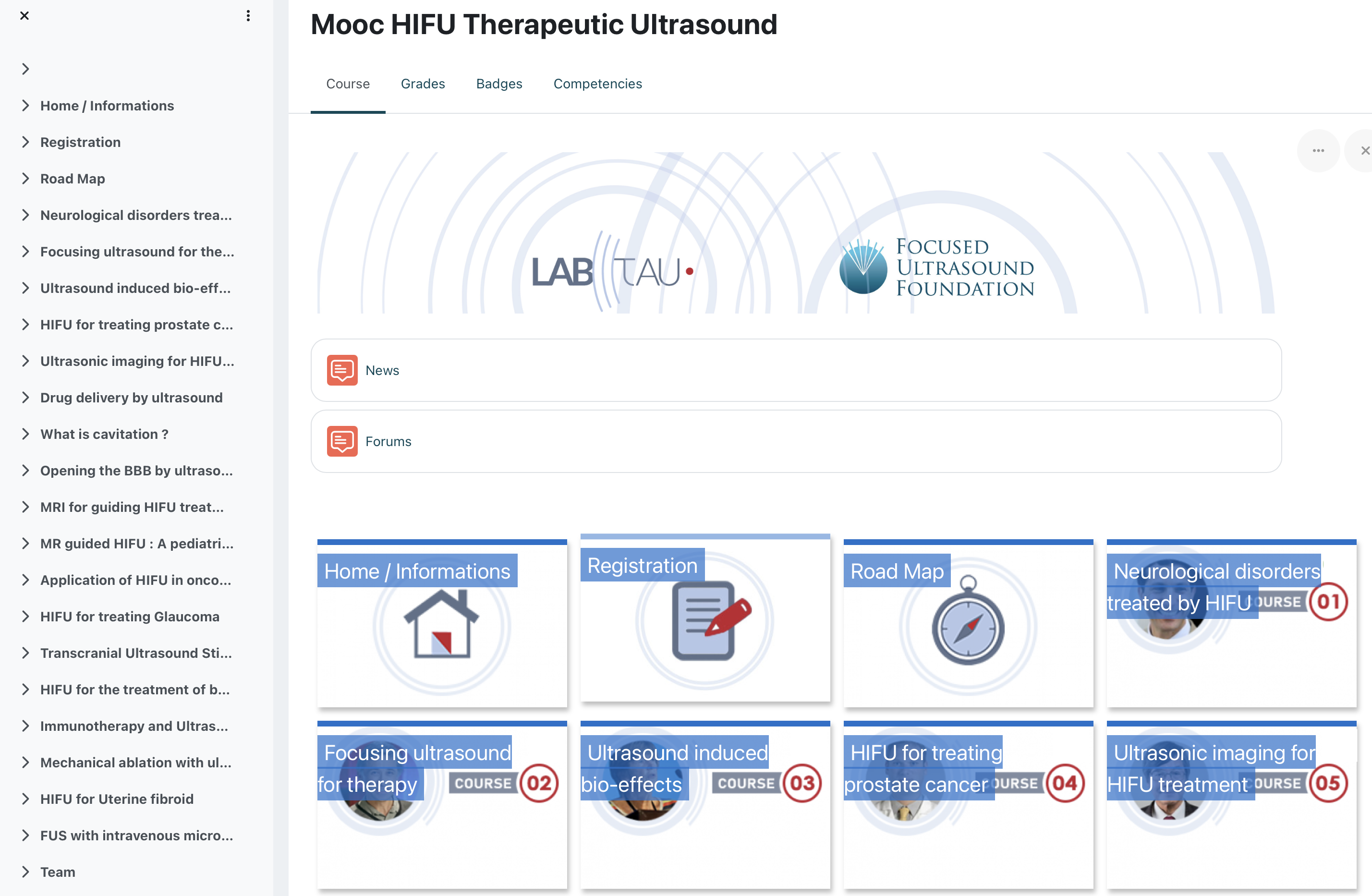
Since September 2023, 18 courses on therapeutic ultrasound are available on the MOOC HIFU supported by the Focused Ultrasound Foundation.
This education program aims at promoting the field of therapeutic ultrasound in its various applications for a targeted audience of Physicians, Students, PhD Students and Patients.
You can register to the MOOC HIFU on the brand new platform using the following link : https://foad.univ-lyon1.fr/course/view.php?id=7
You will find all the details of the connecting procedure on the Registration page , to first “Create your account” and then “Register to the MOOC” to access all the materials available on the platform.
A diploma is also available, if you answer 12 questionnaires selected among the 18 available.
Many thanks to all the speakers and the Focused Ultrasound Foundation for their support.
We hope you’ll enjoy this innovative on line course and that you will contribute to discussions. We are looking forward to your feedback.
Don’t hesitate to relay and follow us both on the LabTau LinkedIn page.
https://www.linkedin.com/company/labtau/
Tuesday 27 May 2025
A travers cette vidéo réalisée par la Fondation pour la Recherche Médicale, Domitille Le Quéré et Axelle Brulport du LabTAU vous présentent un sujet concernant l’amélioration de la prise en charge de l’endométriose : https://www.youtube.com/watch?app=desktop&v=ViWLUYflHtM
Le lien vers cette video est également disponible sur notre chaine Youtube

Thursday 17 April 2025
Author: Martial Defoort
Time: 11H00
Language: French/English
Place: Conference Room at LabTAU
Abstract: Ultrasounds for embedded applications: from power transfer to secured communications.
This presentation will focus on the generation and exploitation of ultrasound produced by piezoelectric transducers ranging from centimeter-scale devices (e.g., ceramic types) to micrometer-scale devices (e.g., PMUTs), operating from the linear to the nonlinear regime. In the first part, the emphasis will be placed on the use of these devices in the linear regime for wireless power transfer, with the goal of efficiently recharging medical implants located deep within the human body. Preliminary results demonstrate that acoustic energy can be transmitted through biological tissues at power levels sufficient to supply devices such as pacemakers, while complying with medical safety requirements.
In the second part, the presentation explores the potential of the nonlinear regime of PMUTs, particularly their ability to generate chaotic signals, for use in acoustic cryptography. By leveraging the intrinsic dynamic complexity of these systems, it becomes possible to envision secure communication schemes between external devices and medical implants, making data exchanges more resistant to interception or hacking. This presentation will highlight the feasibility of generating such chaotic behavior using standard PMUTs, without requiring significant hardware modifications, paving the way for easy integration into existing systems. This original approach, at the crossroads of nonlinear physics, microtechnology, and medical cybersecurity, offers a promising new avenue for securing the next generation of connected implants.
Monday 16 December 2024
Author: Thomas Biscaldi
Time: 14H30
Language: French
Place: Conference Room at LabTAU
Abstract: Hepatocellular carcinoma is a primary tumor of the liver and is estimated to be the fourth leading cause of cancer-related death worldwide. Its incidence has been rising steadily in recent years. The gold standard of treatment is single- or multi-needle interstitial ablation, which aims to destroy the tumor locally. These strategies are proving effective, but have certain shortcomings. Interstitial treatments are not conformal: the shape of the ablation does not adapt to that of the tumor. This is problematic, and can lead to recurrence or serious complications for the patient. Ultrasound technologies could provide a solution in this field, by offering targeted therapies guided byin situ imaging, thus offering a novel modality for the practitioner.
Simulations of a new ultrasonic catheter were carried out to design specifications and verify the feasibility of the project in terms of focusing and ablation. A bimodal ultrasonic catheter with a diameter of 3 mm and 64 piezoelectric elements operating at 5.5 MHz was sub-processed accordingly. First, ultrasound imaging capabilities were assessed and confirmed. The prototype was then characterized electronically and acoustically. The thermal performance of the catheter was studied in three dimensions under MR thermometry, validating the simulation tools and demonstrating the directional aspect of induced heating. These results led to the performance of in vitro tests on animal liver. Centimeter radial ablations confirmed, for the first time, the catheter's ability to perform thermal ablations. The prototype's robustness over the full range of tests was also studied. Finally, the re-installation of an ultrasound navigation platform led to the reconstruction of tumor volumes in 3 dimensions. Combining the catheter with this robotized platform enabled the generation of 3-D volumetric thermal ablations, and the treatment of volumes compatible with primary tumors encountered in clinical practice.
Titre : Ultrasons focalisés interstitiels guidés par la navigation échographique pour les thérapies conformationnelles du carcinome hépatocellulaire
Résumé : Le carcinome hépatocellulaire est une tumeur primaire du foie et est estimé comme étant la quatrième cause de décès liés au cancer dans le monde. Son incidence demeure en constante augmentation ces dernières années. Les traitements de référence sont les ablations interstitielles mono- ou multi- aiguille(s) qui visent à détruire localement la tumeur. Ces stratégies s'avèrent efficaces mais présentent certains défauts. En effet, les traitements interstitiels ne sont pas conformationnels : la forme de l'ablation ne s'adapte pas à celle de la tumeur. Ce phénomène s'avère problématique et peut mener à des récidives ou des complications graves pour le patient. Les technologies ultrasonores pourraient apporter une solution dans ce domaine en proposant des thérapies ciblées guidées par une imagerie in situ offrant ainsi une modalité inédite pour le praticien.
Des simulations d'un nouveau cathéter ultrasonore ont été réalisées pour concevoir un cahier des charges et vérifier la faisabilité du projet en termes de focalisation et d'ablation. Un cathéter ultrasonore bimodal de 3 mm de diamètre et de 64 éléments piézoélectriques fonctionnant à 5,5 MHz a été sous traité en conséquence. Tout d'abord, les capacités d'imagerie échographique ont été évaluées et confirmées. Le prototype a ensuite été caractérisé électroniquement et acoustiquement. Les performances thermiques du cathéter ont été étudiées en trois dimensions sous thermométrie IRM et ont validé les outils de simulation tout en démontrant l'aspect directionnel des échauffements induits. Ces résultats ont conduit à réaliser des essais in vitro sur foie animal. Des ablations radiales centimétriques ont confirmé, pour la première fois, les capacités du cathéter à effectuer des ablations thermiques. La robustesse du prototype sur l'ensemble des essais a été étudiée. Enfin, la remise en place d'une plateforme de navigation échographique a donné lieu à la reconstruction de volumes tumoraux en 3 dimensions. L'association du cathéter avec cette plateforme robotisée a permis de générer des ablations thermiques volumiques en 3 dimensions et de traiter des volumes compatibles avec les tumeurs primaires rencontrées en pratique clinique.

Monday 18 November 2024
Author: Klazina Kooiman
Time: 11H00
Language: English
Place: Conference Room at LabTAU
Abstract: Ultrasound-activated vibrating microbubbles (1-10 µm in size) have shown preclinical potential to boost drug therapy and reduce side-effects for treating cardiovascular disease and cancer because drugs are delivered locally. Recently, safety of the treatment was demonstrated in several clinical trials. Despite the advances in the field, the underlying mechanism of microbubble-mediated drug delivery are poorly understood. One of the reasons for this is the huge range in time scales involved. The time scale of the microbubble vibration is 2 million times per second in a 2 MHz ultrasound field (microseconds), which is many orders of magnitude smaller than the time scale of physiological effects (milliseconds), let alone that of biological effects (seconds to minutes) and clinical relevance (days to months). To allow the investigation of the microbubble-cell-drug interaction at a microsecond and micrometer resolution, unique technology was created by coupling an ultra-high-speed camera (~20 million frames per second recordings) to a custom-built confocal microscope. In this seminar, I will describe new insights gained into the microbubble-cell-drug interaction by using this technology for two different cell types: endothelial cells that line blood vessels and bacteria. For endothelial cells, the focus will be on the microbubble behavior in relation to the drug delivery pathways sonoporation (i.e., cell membrane poration), tunnel formation and cell-cell contact opening, as well as how the cytoskeleton F-actin plays a role. Novel microbubble-mediated treatments for the life-threatening disease bacterial infective endocarditis, either on native heart valves or cardiac devices such as pacemakers, are the focus for the bacteria biofilm work.

Thursday 31 October 2024
Prix Saint Gobain 2023
Le Prix Saint-Gobain 2023 est décerné à Gabrielle Laloy Borgna pour sa thèse intitulée "Micro-élastographie : caractérisation mécanique de la cellule par ondes élastiques" effectuée à l'Université Claude Bernard / Lyon 1 sous la direction de Stefan Catheline.
Son travail porte sur l'imagerie des objets biologiques via la mesure de leurs propriétés mécaniques, une classe de techniques dont la mieux connue est l'échographie. La première partie porte sur une extension de ce type de caractérisations à des échelles nettement plus petites que celles auxquelles elles ont aujourd'hui accès, celle de la cellule vivante unique. Pour cela, elle a mis au point une technique originale pour générer des ondes mécaniques à petite échelle via l'utilisation d'une bulle oscillante. Elle a ensuite tourné son attention vers des amas cellulaires mimant des tumeurs cancéreuses, et a là encore contribué au développement d'une source d'onde innovante à base de nanoparticules magnétiques.
La dernière partie de ce travail porte sur des échelles beaucoup plus grandes, et a trait à l'onde de pouls provoquée par les battements du coeur et qui se propage le long des vaisseaux sanguins. Alors que la communauté scientifique n'avait jusque là connaissance d'un seul type d'onde de pouls, Gabrielle Laloy Borgna en a au cours de sa thèse identifiée une seconde, beaucoup plus lente et impliquant la flexion des vaisseaux sanguins. Ces résultats bouleversent 200 ans d’histoire faisant suite aux travaux de Thomas Young en 1820 et ouvrent des perspectives très prometteuses pour le diagnostic médical (1 Science Advances, 3 APL, 1 brevet).
https://sfpnet.fr/gabrielle-laloy-borgna-et-marlone-vernet-laureats-des-...
Wednesday 30 October 2024
Abstract: The Internationnal Tissue Elasticity Conference (ITEC) was held in Lyon from 21 to 23 October. With a total of around 100 participants, it was a great success and a pleasure to share recent advances in elastography. We look forward to seeing you in Seattle at the end of June 2025. Stefan Catheline, for the organising committee.
Wednesday 16 October 2024
Dr Gabrielle Laloy-Borgna and Dr Stefan Catheline discovered flexural waves in blood vessels, paving the way for more accurate cardiovascular diagnostics.
https://doi.org/10.1126/sciadv.adf1783
This breakthrough could transform heart health assessments!
Learn more: https://scitube.io/dr-gabrielle-laloy-borgna-dr-stefan.../
Tuesday 8 October 2024
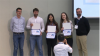
Mr. Thomas BISCALDI, PhD student at the Laboratory of Therapeutic Applications of Ultrasound (LabTAU, French National Institute of Health and Medical Research, University Claude Bernard of Lyon, France), recently received the International Society of Therapeutic Ultrasound's (ISTU) Nadine BARRIE SMITH Award at the 23rd International Symposium for Therapeutic Ultrasound. ISTU's most prestigious student award was established in 2011 in honor of Dr. Nadine BARRIE SMITH's dedication to students in therapeutic ultrasound and recognizes exemplary student achievements at the ISTU Annual Symposium. This fund allows continued recognition of Dr. BARRIE SMITH's legacy as a mentor, and to encourage others to do the same.
The ISTU24 event was held in Taipei (Taiwan) from Sept. 19 to 22, 2024, at the Taipei International Convention Center (TICC) located right in the heart of Taipei City in the international Xinyi business district. Following ISTU tradition, the student award winners were selected by the ISTU Student Awards Committee after pre-selection of abstracts and presentations of their works at either the "Student Award Talks" Session or the "Student Award Posters" Session. Three awards were then given for Best Poster, Best Oral Presentation, and the Nadine BARRIE SMITH Award at the final "Student Award Announcements" session. This year, Mr. Thomas BISCALDI (LabTAU, INSERM, CLB, University Claude Bernard of Lyon, France) shared this session with Ms. Mahsa MOKHLESABADI (Sunnybrook Research Institute, Toronto, Canada) and Ms. Vanessa DREVENAKOVA (Imperial College London, UK) who respectively received the awards for Best Oral and Best Poster Presentations.
Thomas BISCALDI gave a presentation entitled "Dual-Mode US Interstitial Catheter for Robotic US-Navigation-guided Conformal HIFU therapy: in-vitro experimental validation" highlighting their recent contributions to the development of 3D image-guided robot-assisted ultrasound therapy in interventional radiology, in the context of HepatoCellular Carcinoma (HCC) ablation.
Tuesday 10 September 2024
Une de nos doctorante Sybille GREGOIRE est intervenue dans le podcast de l'ASA qui s'appelle Across acoustics (https://www.buzzsprout.com/1537384/15614112) pour expliquer le contenu de son article
Friday 5 July 2024
Lauréats du Prix de Thèse 2023 en GBM
La Société Française de Génie Biologique et Médical et l'Alliance pour le Génie Biologique et Médical, avec le soutien de la Communauté Stic-Santé, ont décerné leurs prix de thèse 2023 à l'occasion des RITS 2024 à l'Université Clermont-Auvergne.
Les quatre finalistes ont présenté leurs travaux lors d'une session spéciale devant le Jury présidé par la Professeur Frédéric Patat, et le choix des membres du Jury a été le suivant :
Le prix Recherche a été décerné à Gabrielle LALOY-BORGNA, pour ses travaux au LabTau (INSERM U1032, Université Lyon1) intitulés "Micro-élastographie : caractérisation mécanique de la cellule par ondes élastiques".
Le prix Innovation a été décerné à Marion TACONNE, pour ses travaux au LTSI (INSERM U642, Université de Rennes) intitulés “Hybrid approach, combining computational and machine-learning models, for the analysis of myocardial strain and cardiac function evaluation”.
Une mention spéciale Recherche a été décernée à Ling HUANG, pour ses travaux au laboratoire Heudiasyc (CNRS UMR 7253, UTC) et à l'Université de Rouen-Normandie intitulés “Medical Image Segmentation with Belief Function Theory and Deep Learning” .
Enfin une mention spéciale Innovation a été décernée à Paul NOBRE, pour ses travaux au Laboratoire CREATIS (CNRS UMR 5220, INSERM U1294, INSA Lyon) intitulés “Sondes et capteurs de champs électromagnétiques à liaisons optiques pour la sécurité en IRM” .
Cette année, le jury était présidé par Frédéric Patat et avait pour membres Olivier Beuf (Responsable d’organisation), Su Ruan, Véronique Migonney et Daniel Racoceanu (SFGBM), Guirec Hillion (AGBM) et Emmanuel Blanc (anciennement CTO EDAP TMS).
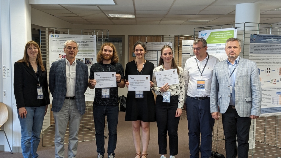
Prix jeunes chercheurs / jeunes chercheuses aux RITS 2024
La SFGBM a récompensé deux jeunes chercheurs lors des journées RITS 2024 qui se sont déroulées à l'Université Clermont-Auvergne.
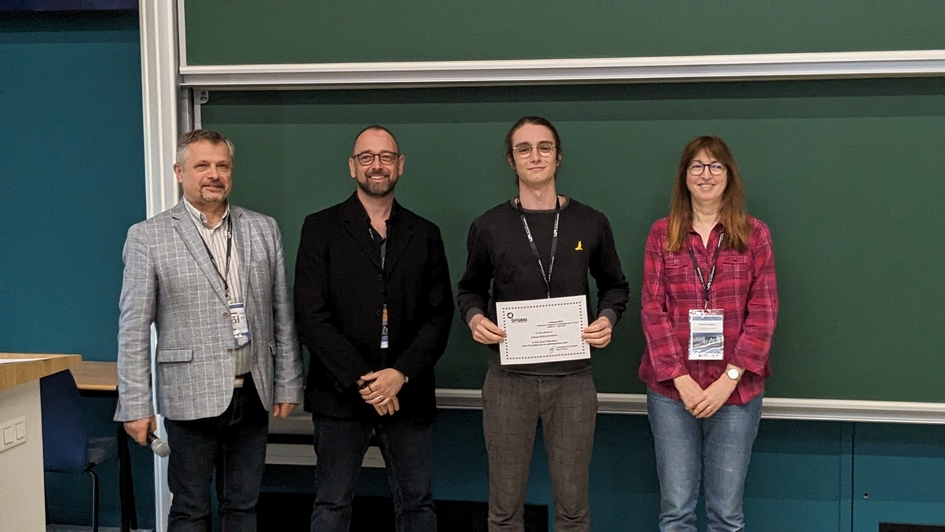
Clément FOULLOUNOUX (LabTAU, INSERM U1032, Centre Léon Bérard, Université Claude Bernard Lyon 1) a reçu le prix Jeune Chercheur de la meilleure communication orale, intitulée "Étude du déclenchement de la cavitation dans le cristallin en vue de traiter la presbytie".
Tom AUBIER (LabTAU, INSERM U1032, Centre Léon Bérard, Université Claude Bernard Lyon 1) a reçu le prix Jeune Chercheur de la meilleure communication affichée, intitulée "In vitro investigation of the biophysical mechanisms underlying ultrasound neurostimulation".

Thursday 30 May 2024
La Fondation de l’Avenir a rencontré Cyril LAFON, docteur en génie biomédical, directeur de recherche en physique à l’INSERM et directeur du LabTAU1 à Lyon, pour qu’il nous présente ses travaux de recherches portant sur l’usage des ultrasons pour soigner le cancer du sein. Ce projet est mené en collaboration avec Marion CORTET, que nous avons également interviewé, notamment sur la dimension clinique en gynécologie de l’application des techniques ultrasonores développées.
L’usage des ultrasons comme traitement thérapeutique, et spécifiquement du cancer, est fréquent en médecine (pour plus de détails, un article de l’INSERM est disponible ici sur le sujet). Il est courant d’utiliser les ultrasons pour traiter par exemple les calculs rénaux, le cancer de la prostate, le glaucome, ou encore les varices des membres inférieurs. Le projet de Cyril LAFON consiste à développer cette technique de traitement pour le cancer du sein, combiné à un traitement par immunothérapie. L’approche sera d’abord testée dans un modèle préclinique de ce cancer, pour ensuite procéder à l’adaptation d’une sonde ultrasonore pour cette partie du corps pour un essai clinique.
Inaudibles à l’oreille humaine, les ultrasons en médecine se montrent particulièrement polyvalents, notamment dans le traitement du cancer. Ils peuvent permettre de détruire les cellules tumorales par la chaleurqu’ils génèrent. En outre, par l’effet d’oscillation mécanique, les ondes ont la capacité de rendre les cellules cancéreuses perméables temporairement aux agents thérapeutiques, et les rendre ainsi plus sensibles à ces molécules. Enfin, les ondes ultrasonores peuvent, en détruisant les cellules tumorales par oscillation de microbulles à leur voisinage, alerter le système immunitaire qui peut plus facilement accéder au cancer et le combattre. Cette action pourrait contribuer à créer un effet de protection durable, s’apparentant à celui offert par la vaccination. C’est cette dernière stratégie, de destruction alertant le système immunitaire, qui a été choisie par Cyril LAFON et son équipe.
____________
1 Laboratoire des applications Thérapeutiques des Ultrasons
A présent, nous vous partageons en image le projet et les explications de Cyril LAFON :

Ce projet fait partie des travaux soutenus par le Fonds Avenir MASFIP. Né en 2014 d’un partenariat entre la Fondation de l’Avenir et la MASFIP (Mutuelle d’Action Sociale des Finances Publiques), le Fonds Avenir MASFIP est dédié à l’amélioration de la compréhension et de la prise en charge des cancers à caractère familial. Son action vise à soutenir directement des équipes de recherche et créer un espace de réflexion sur les enjeux sociaux que pose l'oncogénétique.
Pour plus d’informations sur le projet de Cyril LAFON et sur l’oncogénétique, nous vous invitons à vous rendre sur le site www.oncogenetique.fr

Thursday 23 May 2024
Author: Anam Bhatti
Time: 11H00
Language: English
Place: Conference Room at LabTAU
Abstract:
In response to any cutaneous disease, the morphology and functionality of superficial vasculatures in skin being deteriorated. Superficial vasculatures are characterized as the tortuous and branched structures anastomosed around the diseased tissue. Therefore, visualization of these vessels plays a crucial role in diagnosis and prognosis of cutaneous disorders. Traditionally, high-frequency ultrasound has been used in dermatology for dermal morphological observation, its limited Doppler sensitivity poses challenges for micro-vessels visualization. This study addresses this limitation by employing the ultrafast acquisition techniques of ultrasound and advanced clutter filtering approaches based on singular value decomposition to map the micro-circulation in dermal superficial vasculatures effectively in 2D and 3D imaging.
A novel approach using region-based SVD clutter filtering was proposed to extract the blood flow signals in vasculatures of different dimensions, densities, and flow speed across different layers of the skin. Additionally, a 3D volumetric imaging method was introduced to visualize the superficial vasculatures from various perspectives, using continuous mechanical translation to scan the imaging area and the section-wise region-based SVD processing was proposed to extracts the blood flow signal efficiently. Post-processing algorithms such as top-hat transform morphological filter and non-local means filter were implemented to remove the noises and enhance the image quality.
The effectiveness of the proposed approaches was evaluated using custom-designed micro-flow phantoms mimicking different types of superficial micro-vessels. In-vivo experiments on healthy subject demonstrated the visualization of micro-vessels with a minimum diameter of 51 μm in the dermal layers. Acquired in-vitro and in-vivo results highlights the efficacy of the proposed 2D and 3D approaches to visualize the superficial micro-vessels for an effective diagnosis of cutaneous diseases.

Thursday 2 May 2024
Author: José MC CORTES & Inas H FASRIS
Time: 11H00
Language: English
Place: Conference Room at LabTAU
Abstract: #1 MECHANICAL CHARACTERISATION OF THE TUMOUR MICROENVIRONMENT BY MICROELASTOGRAPHY This workoutlines the mechanical characterization of the tumor microenvironment using microelastography as part of a broader research project focused on modifying the response of tumor cell microenvironments. The overarching objective is to induce therapeutic effects in various tumors through controlled mechanical shear waves. Key components of the project include a theoretical framework for analyzing mechanically induced signaling pathways in tumor progression and treatment, the development of a transducer to generate and insonate mechanical shear waves in a controlled microenvironment, and the design of a dynamic optical microelastography technique to characterize the induced mechanical microenvironment and tumors. Specifically, this thesis focuses on the design of dynamicopticalmicroelastography to provide experimental data for computational models and verify the functionality of the bioreactor. The main objectives include a parametric study of a biocompatible matrix to create a microenvironment for tumor spheroids, mechanical studies of tumor spheroids and their microenvironments, analysis of viscoelastic models and parameters of the matrix and tumor, and the investigation of in vitro tumor growth at the mechanical level and its impact on the microenvironment. This work aims to deepen understanding of tumor mechanobiology and contribute to the development of novel therapeutic approaches.
#2:UNVEILING TISSUE SECRETS: TRANSITIONING THROUGH TORSIONAL WAVE ELASTOGRAPHY TO EXPLORE THE IMPACT OF TISSUE VASCULARITY ON VISCOELASTICITY VALUES
Part I: Torsional Wave Elastography (TWE) Sensor Design and Application on soft tissue
The first part of the presentation pivots to the practical application of these theoretical insights, focusing on the design and clinical application of a Torsional Wave Elastography (TWE) sensor. This innovative tool represents a leap forward in non-invasive cervical and skin lesion diagnostics among others, moving beyond conventional elasticity metrics to capture a fuller spectrum of mechanical biomarkers, including viscosity, anisotropy, and heterogeneity. Through detailed analysis and clinical validation, we demonstrate how TWE can significantly enhance diagnostic accuracy, particularly in identifying and characterizing skin lesions.
Part II: Bridging Vascularity and Mechanics
This research project, "Mapping the Vascular Maze: Investigating the Interplay Between Tissue Vascularity and Viscoelasticity," conducted at the University of Granada, aims to uncover the complex relationship between the structure of blood vessels within tissues and their ability to deform and recover, known as viscoelasticity. Centredaround the fractional alpha (α) parameter in viscoelastic models, this study explores how vascularity influences tissue mechanics, with a particular focus on cancer where abnormal blood vessel patterns are prevalent. By combining advanced computational models with experimental work using 3D-printed tissue phantoms, the team seeks to link tissue vascularity with mechanical properties, understand vascular changes in tumours, and improve predictive models for tissue behaviourunder stress. This interdisciplinary effort integrates physics, biology, engineering, and materials science, aiming to enhance diagnostics and treatment strategies by leveraging the mechanical properties influenced by tissue vascularity.

Thursday 22 February 2024
Author: Jessica Gannon
Time: 11H00
Language: English
Place: Conference Room at LabTAU
Abstract:
Ultrasound-Guided Non-Invasive Pancreas Ablation Using Histotripsy: Feasibility Studies in Porcine Models
Pancreatic cancer is an aggressive disease that is typically diagnosed late-stage leading to poor prognosis and overall survival. Even with advances in treatment, the five-year survival rate for patients is only 12.5% with only 20% of patients presenting with resectable tumors. Histotripsy is a non-invasive, non-thermal, and non-ionizing focused ultrasound ablation method that has recently been FDA approved for treating hepatic tumors and is currently being investigated for other malignancies. This study expands upon prior work by our group investigating the feasibility of non-invasive pancreas ablation using ultrasound-guided histotripsy in a healthy chronic porcine model. Additional work by our group includes the development of an immunocompromised porcine model allowing us to surgically inject and grow human Panc-01 tumors orthotopically to target with histotripsy. While results from these studies are promising, they also highlight the challenges of targeting the pancreas through an extracorporeal approach due to overlying gas-filled tissues. Therefore, we are also investigating the development of endoscopic histotripsy devices for minimally invasive tissue ablation which has led to the collaboration between our two labs.
Thursday 15 February 2024
Author: Marie Poulain-Zarcos
Time: 14H00
Language: English
Place: Meeting Room at LabTAU
Abstract: Microstructure of sheared dense suspensions of Red Blood Cells (RBCs)
Blood is a non-Newtonian fluid that exhibits shear-thinning behavior (decreasing viscosity with increasing shear rate). This increases oxygen transport in the body. This shear-thinning behavior is influenced by (1) blood aggregation and (2) RBC deformability. Both of these characteristics can be affected in certain diseases such as diabetes or sickle-cell anemia (hyper-aggregation and greater rigidity of RBCs, which can lead to painful vaso-occlusive crises). In this seminar, we propose two experimental techniques for assessing these two characteristics.
Firstly, using ultrasonic measurements, I will present the characterization of blood aggregation (aggregate size, polydispersity, etc.). The clinical application is to estimate the rate of aggregation in blood and (in the longer term) to enable pre-diagnosis.
Secondly, using optical measurements in a microfluidic channel, I will present the modification of RBC microstructure and dynamics induced by a change in their deformability. A comparison with sickle cell cells will be presented. The clinical application is to estimate the variability of RBC deformability and (in the longer term) to enable monitoring of sickle cell disease.

Thursday 25 January 2024
Author: Adrien Rohfritsch
Time: 11H00
Language: French/English
Place: Conference Room at LabTAU
Abstract:
La connaissance précise des propriétés acoustiques d’un milieu hétérogène est d’une importance majeure dans de nombreux domaines d’application, tels que l’imagerie, le contrôle non destructif, le développement de métamatériaux ou la thérapie par ultrasons focalisés. La propagation des ultrasons dans un tel milieu est régie à la fois par les propriétés de l’onde incidente, la taille des hétérogénéités du milieu, leurs propriétés acoustiques et leur distribution spatiale. La prise en compte de tous ces facteurs est un défi qui limite la plupart du temps le champ d’application d’une ´étude numérique ou d’un modèle théorique. Nous présentons un code de calcul récent, MuScat, qui permet de pallier ces problèmes. Il rend possible le calcul du champ formé par l’interaction de l’onde incidente avec un milieu peuplé de plusieurs milliers de diffuseurs cylindriques ou sphériques. Ce code fréquentiel prend en compte les effets de diffusion multiple `a tous les ordres ainsi que la taille des particules, leurs propriétés mécaniques et leurs positions. Ce dernier point permet de modéliser exactement n’importe quel type de microstructure (i.e. la distribution spatiale des diffuseurs dans l’espace). La diffusion par une particule est modélisée par la matrice de diffusion décomposée sur la base modale, et les interactions entre particules sont décrites à l’aide de la théorie de la diffusion multiple linéaire.
Une première étude d´démontre la possibilité de tirer profit des corrélations spatiales à courte portée (appelées SRC pour Short Range Correlation) et longue portée (SHU, pour Stealth Hyperuniform) afin de fabriquer des matériaux aux propriétés surprenantes et très prometteuses pour le d´développement de m´métamatériaux de nouvelle génération. Une seconde ´étude se focalise sur la détection de lésions créées par ultrasons focalisés de haute intensité (HIFU) à l’intérieur du tissu hépatique. Le passage du faisceau focalisé créé une anisotropie locale dans le réseau cellulaire, qui modifie le signal rétrodiffusé par le milieu. Nous quantifions ce phénomène grâce à des simulations numériques et le corrélons à l´évolution du facteur de structure de la zone traitée. Nous montrons pour finir de premiers résultats expérimentaux ouvrant la voie à un nouveau moyen de suivi temps réel des traitements thermiques par HIFU.

Thursday 11 January 2024
Author: Juliette Pierre
Time: 11H00
Language: French
Place: Conference Room at LabTAU
Abstract: Acoustic signature of hydrodynamics events
Gas-liquid systems are ubiquitous in industrial, food, biological or geophysical contexts. The popping noise of a bursting bubble, the crackle sounds of ageing foams, the whistling of nucleating water, the fizzing of champagne, the drumming of rain and the thud of degassing volcano magmas evidence the radiation of sound by these violent interfacial hydrodynamic events.
Such events evolve according to various processes occurring at several different length scales and over several different time scales. These non-equilibrium systems are mainly studied using fast optical imaging, which allows us to follow the reconfiguration of certain liquid-gas interfaces over time. By adding acoustic measurements, we also have access to the pressure field generated.
In this presentation I will present our recent investigations about the spatio-temporal acoustical signature of violent hydrodynamic events such as : bubble bursting, drop impact or film bursting. By combining the use of acoustic sensors, fast optical imaging and theoretical modeling, we aim to monitor and characterise these violent events and elucidate the physical mechanisms at play in their existence and their evolution.

Wednesday 22 November 2023
Author: Myléva DAHAN
Time: 2 PM
Language: French
Place: Conference Room at LabTAU
Résumé:
L'utilisation des ultrasons thérapeutiques, de l'immunothérapie, et de la délivrance de médicaments via des nanoparticules représente une avancée significative dans le domaine de la recherche en oncologie. Chacune de ces modalités thérapeutiques offre des avantages distincts pour le traitement du cancer, mais leur combinaison promet une approche révolutionnaire pour lutter contre cette maladie dévastatrice. En combinant plusieurs modalités, nous visons à créer une réponse thérapeutique améliorée et synergique pour lutter contre le cancer. Cette approche polyvalente tire parti des avantages spécifiques de chaque méthode pour obtenir des résultats plus efficaces. La conception et la caractérisation de l’appareil ultrasonore confocal multimodal dédié aux applications pré-cliniques a fait l’objet d’un premier travail. Cette plateforme offre une polyvalence pour différentes modalités de traitement préclinique, une robustesse et une stabilité permettant la personnalisation et la facilité de comparaison de différentes approches thérapeutiques ultrasonores,
telles que la cavitation inertielle, l’histotripsie et l’ablation thermique.
Ensuite, dans une seconde partie, des nanoparticules à base de liposome et complexées à un agent immunostimulant ont été développées afin de booster le système immunitaire. Ces nanoparticules, capables de complexer l'agoniste TLR3 PolyIC, se sont révélées non cytotoxiques et stables, préservant l'activité du PolyIC et peuvent activer efficacement les cellules dendritiques, offrant des opportunités de combinaisons thérapeutiques.
Enfin, dans une dernière partie, trois combinaisons distinctes ont été examinées sur des modèles pré-cliniques : (1) l'utilisation d'ultrasons mécaniques en conjonction avec des nanoparticules, (2) l'application d'ultrasons thermiques combinée à un inhibiteur de point de contrôle, à savoir l'anti-PD1, et (3) l'association d'ultrasons mécaniques, d'un inhibiteur de point de contrôle (anti-PD1) et d'une hormonothérapie à base de Tamoxifène, chacune présentant ses avantages et inconvénients. Des pistes d'amélioration sont également proposées pour ces approches combinées.
Title: « Therapeutic ultrasound for oncology applications : Optimization of thermal and mechanical therapies with a confocal system for combined treatments, and development of nanoparticles for immune activation »
Abstract:
The use of therapeutic ultrasound, immunotherapy, and drug delivery via nanoparticles represents a significant advancement in the field of oncology research. Each of these therapeutic modalities offers distinct advantages for cancer treatment, but their combination promises a revolutionary approach to combat this devastating disease. By combining different modalities, our goal is to create an enhanced and synergistic therapeutic response to fight cancer. This versatile approach leverages the specific benefits of each method to achieve more effective results. The design and characterization of a multimodal confocal ultrasound device dedicated to preclinical applications were the focus of the initial work. This platform provides versatility for various preclinical treatment modalities, robustness, and stability, allowing for customization and easy comparison of different ultrasound therapeutic approaches, including inertial cavitation, histotripsy and thermal ablation.
In the second part, nanoparticles based on liposomes and complexed with an immunostimulant agent were developed to boost the immune system. These nanoparticles, capable of complexing the TLR3 agonist PolyIC, have proven to be non- cytotoxic and stable, preserving the activity of PolyIC. They can effectively activate dendritic cells, providing opportunities for therapeutic combinations.
Finally, in the last part, three distinct combinations were examined in preclinical models: (1) the use of mechanical ultrasound in conjunction with nanoparticles, (2) the application of thermal ultrasound combined with a checkpoint inhibitor, namely anti- PD1, and (3) the combination of mechanical ultrasound, a checkpoint inhibitor (anti- PD1), and Tamoxifen-based hormonal therapy, each presenting its own advantages and disadvantages. Suggestions for improvement are also proposed for these combined approaches.

Friday 17 November 2023
Author: Alice Ganeau
Time: 2 PM
Language: French
Place: Conference Room at LabTAU
Abstract:
La presbytie est un défaut visuel qui touche la majorité de la population à partir de 45-50 ans. En effet, cette perte de la capacité à visualiser de près résulte du vieillissement naturel du cristallin. Les corrections optiques sont les solutions les plus couramment utilisées pour améliorer la vision de près
des patients presbytes. Il existe également des chirurgies correctrices qui remodèlent la cornée ou, de manière plus radicale, remplacent le cristallin par une prothèse. Cependant, aucune de ces solutions ne ciblent directement la source de la presbytie, qui est la rigidification du cristallin. L’une des causes biologiques de ce processus serait liée à l’agrégation des protéines du cristallin. Les ultrasons focalisés de haute intensité, en particulier la cavitation ultrasonore, pourraient offrir une nouvelle approche pour le traitement de la presbytie, en détruisant mécaniquement ces agrégats. Cette thèse étudiera donc la faisabilité d’utiliser l’activité de cavitation pour un potentiel traitement de la presbytie. Le deuxième objectif est de développer une méthode d’élastographie ultrasonore par ondes de surface, pour mesurer l’élasticité du cristallin. En effet, l’absence de diffuseurs ultrasonores à l’intérieur de celui-ci contraint l’utilisation des ondes se propageant à sa surface. Cette méthode sera appliquée sur une large gamme de fréquences afin d’observer d’éventuels effets de guidage en raison de la petite taille de cet organe, qui est inférieure au centimètre.
Dans un premier temps, des études de faisabilité, visant à initier un nuage de cavitation ultrasonore au sein d’échantillons de cristallins porcins ex vivo, ont été mises en place. Un dispositif ultrasonore dont la forme et la taille sont compatibles avec celles d’un globe oculaire a permis de déclencher de manière répétable des nuages de cavitation. Plusieurs modalités d’imagerie ont été mises en place pour suivre en temps réel le traitement. L’imagerie échographique a permis de visualiser la formation des nuages de bulles et leur dynamique de dissolution après traitement. La cartographie de l’activité de cavitation a pu être réalisée à l’aide de méthodes d’imagerie passive. Ultérieurement, les effets sur la structure interne du cristallin ont pu être observées microscopiquement à l’aide d’un protocole de coupes histologiques. Ces résultats ont montré la nécessité de développer une méthode pour suivre en temps réel le traitement ultrasonore du cristallin, et plus particulièrement pour monitorer les changements d’élasticité.
Dans un second temps, les algorithmes d’élastographie par corrélation de bruit ont été évalués pour détecter un ramollissement induit par la cavitation ultrasonore dans un gel mimant l’agrégation des protéines du cristallin. Puis, l’adaptation de ces algorithmes a permis de mesurer avec précision la dispersion des ondes de surface sur une large gamme de fréquence. Différents régimes de propagation des ondes de surface ont pu alors être observés. La méthode a été appliquée sur des gels plans de gélatine et d’agarose, des inclusions numériques et des échantillons de cristallin porcins excisés de l’œil. L'étude de la dispersion des ondes a montré la nécessité de travailler avec des fréquences suffisamment élevées pour ne pas dépendre des limites du milieu, susceptibles de guider les ondes. Les propriétés viscoélastiques du cristallin ont pu alors être quantifiées. Cependant, à ces fréquences, il n’a pas été possible de détecter de changements locaux d’élasticité en profondeur du cristallin. En effet, il semble que la membrane du cristallin sur laquelle se propage les ondes puisse influencer leur vitesse de propagation, perturbant la mesure.
Ces travaux de thèse représentent les premiers résultats de faisabilité d’initiation de cavitation ultrasonore dans le cristallin à l’aide d’un dispositif cliniquement compatible pour le développement d’un potentiel traitement de la presbytie. De même, les études d’élastographie ont permis de poser de premières pistes de développement de méthodes pour suivre le traitement, qui permettra à termes d’évaluer l’efficacité d’une telle thérapie.

Thursday 12 October 2023
Jeudi 12 octobre le LabTAU a eu l'honneur d’accueillir M. Didier Samuel (Président Directeur Général de l’Inserm) accompagné de M. Frédéric Fleury (Président de l’Université Lyon 1) pour une présentation des différentes activités du Laboratoire.
Ce fut l’occasion d’une démonstration par nos chercheurs de l’utilisation des ultrasons à la thérapie.
Friday 21 April 2023
Place: Lyon (Palais de la Bourse) - Satellite of ISTU/EUFUS 2023
It is with great enthusiasm that we announce the arrival of the 2nd edition of HIFUture which will be held following the ISTU/EUFUS 2023 international symposium, at the Palais de la Bourse in Lyon, on Friday April 21, 2023.
LabTAU and the Hospices Civils de Lyon invite you to this day dedicated to meetings between clinicians, engineers and industrials involved in ultrasound treatments in France and abroad.
The symposium program will include presentations by clinical and academic leaders on : i) a prospective of therapeutic ultrasound with a special focus on the future of HIFU in Interventional Radiology; ii) the physics of ultrasound and the key clinical applications of this technology; iii) the latest clinical innovations and those to come in various medical specialties.
The event will take place in person and can also be followed remotely (virtual part). After the success of the 2021 edition, we are expecting you on April 21, 2023 for this 2nd edition.
To participate to HIFUture 2023 : HIFUture2023
Organizers : Gil Dubernard & Cyril Lafon. Secretaries : Charles-André Philip & W. Apoutou N’Djin
Monday 17 April 2023
ISTU – EUFUS 2023 Lyon (France) April 17th to 20th
Meet us in Lyon for the second joint meeting of the International Society for Therapeutic Ultrasound (ISTU) and the European Focused Ultrasound Charitable Society (EUFUS)
Register now : https://istu.org/

Thursday 2 February 2023
Author: Christina Tsarsitalidou
Time: 11H00
Language: French/English
Place: Conference Room at LabTAU
Abstract: Seismic imaging constitutes a fundamental building block of Earth Science research as the obtained
images or snapshots of our planet’s interior challenge our understanding of plate tectonics, yield
information on fault zone environments and inform many near-surface engineering applications. In
this work, we explore the imaging potential of refocusing surface waves reconstructed from noise
correlation functions so as to estimate the seismic velocity. At zero lag-time, converging surface
wave fields create a large amplitude feature at the origin referred to as focal spot. Its properties are
governed by local medium properties and have long been used in medical imaging approaches such
as passive elastography. We treat dense seismic arrays equivalent to medical ultrasound transducers
that allow the reconstruction of focal spots at near-field distances. We focus on demonstrating the
general applicability of the method using the USArray dataset, but we also investigate the biasing
effects of ambient wave field properties on our estimates, like impinging body wave energy and
non-isotropic energy flux.

Tuesday 13 December 2022
Author: Sophie Cambronero
Time: 14h00
Language: French
Place: Conference Room at LabTAU / Visio Teams
Abstract: Les ultrasons focalisés de haute intensité (HIFU) sont une modalité de traitement non invasive des tumeurs solides qui permet une destruction tissulaire localisée et en profondeur des tissus. Les travaux de ce manuscrit décrivent le développement de stratégies de traitement et la conception d’un transducteur de géométrie torique dédié au traitement non invasif des tumeurs hépatiques. Dans un premier temps, un dispositif
HIFU torique existant et conçu pour le traitement peropératoire des tumeurs hépatiques a été utilisé afin d’élargir les perspectives de traitement peropératoire pour les tumeurs localisées au confluent cavo-sus- hépatique. Cette approche de traitement a été évaluée sur une étude préclinique et a montré la faisabilité de la procédure et la tolérance à moyen du traitement. Ce même dispositif a ensuite été utilisé pour déterminer la faisabilité d’un traitement HIFU complètement non invasif avec un transducteur de géométrie torique. La faisabilité et l’innocuité de ce traitement non invasif a été démontré sur une étude préclinique in vivo chez le modèle porcin. Ces travaux ont mis en lumière la nécessité d’une personnalisation des traitements HIFU non invasif afin d’épargner les tissus intermédiaires sains. Sur la base de ces expériences, un nouveau système de ciblage a été développer afin d’assurer un positionnement précis de l’axe HIFU avec un intérêt particulier pour les traitements réalisés proches de structures à risques. Les performances de nouveau système ont été démontrées sur deux protocoles in vivo pour le traitement du STT (Syndrome Transfuseur Transfusé) et des tumeurs du trigone vésical. Un nouveau dispositif de traitement HIFU a ensuite été conçu et développé pour le traitement non invasif des tumeurs hépatiques. La découpe spécifique de ce transducteur torique tronqué permet un élargissement du volume traité sans déplacement mécanique du transducteur. Les performances thérapeutiques de ce dispositif ont été évaluées sur des essais in vitro et in vivo chez le modèle porcin. Ces travaux représentent la première brique d’un traitement non invasif des tumeurs hépatiques avec un transducteur HIFU torique.

Monday 12 December 2022
Author: Dr Laura CURIEL - Assistant Professor - Department of Electrical and Software Engineering University of Calgary
Time: 11H00
Language: French
Place: Conference Room at LabTAU
Abstract: Le pilotage multiaxial est une nouvelle méthode d’excitation des matériaux piézoélectriques qui utilise une combination des modes de vibration extensionnel et latéral pour moduler le faisceau ultrasonore produit. Il a été démontré que cette méthode peut produire différents effets : une modulation de l’efficacité électroacoustique, une déflection de l’axe d’émission sans avoir à utiliser multiples éléments piézoélectriques, et un control et réduction de la taille focale, en particulier quand un réseau d’éléments multiaxial est utilisé. Dans ce séminaire on discutera les principes du pilotage multiaxial des transducteurs, ses avantages et les applications initiales explorées par notre groupe de recherche. Ensuite, une introduction sur les applications émergentes de la méthode en ce qui concerne des dispositives animaux, ou la réduction de la taille focale présente des avantages, seront décrites.

Friday 9 December 2022
Author: Audrey SIVADON - PhD Student at LabTAU - Inserm U1032
Time: 9H30 AM
Language: French
Place: LabTAU Conference Room / Visio Microsoft Teams
Abstract: L’imagerie passive s’appuie sur des algorithmes de formation de voie qui nécessitent des sondes de grande ouverture pour offrir de bonnes résolutions axiales ; or, en imagerie passive 3D, les sondes matricielles actuellement commercialisées ne vérifient pas cette contrainte. De plus, ces sondes présentent un grand nombre d’éléments, ce qui rend leur utilisation particulièrement lourde. Ce travail de thèse porte sur l’étude et l’amélioration de l’imagerie passive de la cavitation en s’intéressant à deux aspects en particulier : (i) la mise en œuvre pratique et efficace de l’imagerie passive en 3D, (ii) la problématique de l’imagerie de sources étendues telles que des nuages de cavitation.
Nous avons combiné l’application des méthodes sparse (pour réduire le nombre d’éléments actifs de la sonde utilisée) et la transposition du 2D au 3D des algorithmes adaptatifs dans le domaine fréquentiel. Ce formalisme utilise l’estimation robuste de la matrice de densité inter-spectrale (CSM) et nous a permis d’implémenter simplement et efficacement différents algorithmes : Delay-And-Sum (DAS), Robust-Capon-Beamformer et Pisarenko. L’efficacité de ces algorithmes en 3D a été testée en termes de largeur à mi-hauteur, de contraste et d’erreur de position, sur une source ponctuelle en simulations et sur une expérience de réflecteur ponctuel. Enfin, pour répondre à la réalité des nuages de cavitation, nous nous sommes intéressés au comportement de ces méthodes de reconstruction dans le cas de sources étendues. Nos simulations en 2D montrent l’évolution des images reconstruites en fonction de caractéristiques du nuage de cavitation.
Ce travail apporte une solution concrète de mise en œuvre simple de l’imagerie passive 3D ainsi que des éléments de réponse quant aux attentes sur la localisation et la caractérisation d’un nuage de cavitation.
Thursday 7 July 2022
Author: Tristan Jaouen
Time: 14H00
Language: French
Place: LabTAU Conference Room / Visio Microsoft Teams
Link: https://teams.microsoft.com/l/meetup-join/19%3ameeting_YTcwOTlhMTEtOWU1OS00YjVkLTg1MzktZjRmZmE5MWRlYzAy%40thread.v2/0?context=%7b%22Tid%22%3a%2299dde8fb-92f6-414d-bc5e-44ffa3e2419c%22%2c%22Oid%22%3a%2206fbf2f7-63eb-4935-8487-1fe7674caef4%22%7d
Abstract:
We developed a region of interest-based (ROIs) computer-aided diagnosis system (CAD) to characterize International Society of Urological Pathology grade (ISUP) ≥2 prostate cancers at multiparametric MRI (mp-MRI). Image parameters from two multi-vendor datasets of 265 pre-prostatectomy and 112 pre-biopsy MRIs were combined using logistic regression. The best models used the ADC 2nd percentile (ADC2) and normalized wash-in rate (WI) in the peripheral zone (PZ) and the ADC 25th percentile (ADC25) in the transition zone (TZ). They were combined in the CAD system.
The CAD was retrospectively assessed on two multi-vendor datasets containing respectively 158 and 105 pre-biopsy MRIs from our institution (internal test dataset) and another institution (external test dataset). Two radiologists independently outlined lesions targeted at biopsy. The Prostate Imaging-Reporting and Data System version 2 (PI-RADSv2) score prospectively assigned at biopsy and the CAD score were compared to biopsy findings. At patient level, the areas under the Receiver Operating Characteristic curve (AUC) of the PI-RADSv2 score were 82% (95% CI: 74-87) and 85% (95% CI: 79-91) in the internal and external test datasets respectively. For both radiologists, the CAD score had similar AUC results in the internal (82%, 95% CI: 76-89, p=1; 84%, 95% CI: 78-91, p=1) and external (82%, 95% CI: 76-89, p=0.82; 86%, 95% CI: 79-93, p=1) test datasets. Combining PI-RADSv2 and CAD findings could have avoided 41-52% of biopsies while missing 6-10% of ISUP≥2 cancers.
The CAD system confirmed its robustness showing good discrimination of ISUP ≥2 cancers in a multicentric study involving 22 different scanners with highly heterogeneous image protocols. In per patient analysis, the CAD and the PI-RADSv2 had similar AUC values (76%, 95% CI: 70-82 vs 79%, 95% CI: 73-86; p=0.34) and sensitivities (86%, 95% CI: 76-96 vs 89%, 95% CI: 79-98 for PI-RADSv2 ≥4). The specificity of the CAD (62%, 95% CI: 53-70 vs 49%, 95% CI: 39-59 for PI-RADSv2 ≥4) could be used to complement the PI-RADSv2 score and potentially avoid 50% of biopsies, while missing 13% of ISUP ≥2 cancers. These findings were very similar to those reported in the single center test cohorts. Given its robustness, the CAD could then be exploited in more specific applications.
The CAD first provided good discrimination of ISUP ≥2 cancers in patients under Active Surveillance. Its AUC (80%, 95% CI: 74-86) was similar to that of the PI-RADS score prospectively assigned by specialized uro-radiologists at the time of biopsy (81%, 95% CI: 74-87; p=0.96). After dichotomization, the CAD was more specific than the PI-RADS ≥3 (p<0.001) and the PI-RADS ≥4 scores (p<0.001). It could offer a solution to select patients who could safely avoid confirmatory or follow-up biopsy during Active Surveillance (25%), while missing 5% of ISUP≥2 cancers.
Finally, the CAD was tested with the pre-prostatectomy mp-MRIs of 56 Japanese patients, from a population which is geographically distant from its training population and which is of interest because of its low prostate cancer incidence and mortality. The CAD obtained an AUC similar to the PI-RADSv2 score assigned by an experience radiologist in the PZ (80%, 95% CI: 71-90 vs 80%, 95% CI: 71-89; p=0.886) and in the TZ (79%, 95% CI: 66-90 vs 93%, 95%CI: 82-96; p=0.051).
These promising and robust results across heterogeneous datasets suggest that the CAD could be used in clinical routine as a second opinion reader to help select the patients who could safely avoid biopsy. This CAD may assist less experience readers in the characterization of prostate lesions.

Monday 16 May 2022
Author: Laurent Sandrin
Time: 11H00
Language: French/English
Place: Conference Room at LabTAU
Abstract:
Premier dispositif commercial d’élastographie impulsionnelle, FibroScan (Echosens, Paris) a initialement été développé en 2001-2003 pour mesurer l’élasticité du foie afin de déterminer le stade de fibrose des patients atteints de maladie chronique du foie. Depuis 2011, FibroScan mesure également l’atténuation ultrasonore pour quantifier la stéatose hépatique. Avec plus de 7000 dispositifs utilisés en recherche et en routine clinique dans la grande majorité des départements d’hépatologie dans le monde, FibroScan a contribué à transformer la pratique de l’hépatologie en apportant une alternative simple, rapide et non-invasive à la biopsie hépatique.
Monday 2 May 2022
Author: Tristan Deruelle
Time: 13h30
Language: English
Place: Conference Room at LabTAU and video (need registration by clicking the button above)
Abstract:
Prostate cancer is the second most prevalent cancer in men worldwide. It is suspected when the PSA density is high or/and the superficial prostate feels hard during digital rectal examination. Multiparametric MRI is now recommended prior biopsy when detecting for cancer. However, image interpretation is challenging, even for specialists, and brings many false-positive. Elastography is a technique to assess tissue stiffness by inducing small vibrations. It could provide a 3D map of the stiffness of the prostate. We believe that MR elastography could complement the current multiparametric MRI. Given prostate location and consitution, wave propagation is difficult though. The current work presents the design of a non-invasive wave generation device for the prostate. Then, a new field separation algorithm is presented. This algorithm provides a better estimation of the stiffness, and the correction of artefact generated by common vibrators. Finally, this algorithm can have applications in porous media. Indeed, in poro-elastic materials, a slow compression wave propagates. We observe such a wave in an agar gel, in a foam phantom, and in vivo in human kidney graft. In addition to the classic shear wave velocity estimation, it is now possible to estimate the compression wave velocity. This is an additional piece of information that the operator can use in its diagnostic. In the future, more porous parameters could be derived.
Saturday 26 March 2022
Les équipes du LabTAU participaient au 5ème Ekiden (Marathon par équipe de 6).
En plus du soleil, au programme pour nos coureurs: Bonne humeur, esprit d’équipe, bien être, performance, enthousiasme et solidarité.
Bravo à tous!

Tuesday 22 February 2022
Author: Jérôme Sallet & Clément Hebert
Time: 11H00
Language: French/English
Place: Conference Room at LabTAU
Abstract: Part 1: The development of wireless and battery free sensors for biomedical applications is a fast growing research and industrial field. It promises to greatly improve the patient’s comfort during the diagnosis but also in the treatment of chronic diseases. While the standard technologies have been based for the last decade on electromagnetic waves, ultrasonic powering and communication is a very interesting way to reduce the size of the sensor in order to develop minimally invasive implantable technology. In this context, the Wireless Neural Interface team of the Grenoble Institute for Neuroscience is developing a new type of ultrasound-based sensor for biosensing and neural recording. The presentation will show the mm-sized prototype that was used to establish the transfer curve that links the amplitude of the Echo signal to an electric potential in a physiological solution. This curve represents the first step toward real time recording.
Part 2: Abstract: Transcranial ultrasonic stimulation (TUS) is an emerging method whereby low-intensity ultrasound is delivered through the skull to brain tissue resulting in reversible disruption of neuronal activity at the targeted site. Although the exact mechanisms by which ultrasound effects neuromodulation are not fully characterized, the goal of this presentation is to show that the technique is safe and can be used to modulate brain activity and behaviour with a good anatomical precision. TUS neuromodulatory effects were measured by examining relationships between activity in each targeted area and the rest of the brain using resting-state functional magnetic resonance imaging (fMRI) collected under anaesthesia. Importantly those targeted regions could either be superficial cortical areas (e.g. medial premotor, frontopolar cortex), or deep subcortical structures (e.g. Amygdala, Hippocampus, Basal Forebrain). With the specific protocol used, dissociable and focal effects on neural activity could not be explained by auditory confounds. Furthermore, offline effects were shown to last for more than two hours post-stimulation. With such long lasting effect, we were able to test in separate experiments for the specific contribution of perigenual cingulate cortex to counterfactual reasoning, of the lateral orbitofrontal cortex to credit assignment and of the mid-superior temporal sulcus to computing social prediction in primates.

Tuesday 1 February 2022
Author: Elodie Cao
Time: 15h
Language: French
Place: Conference Room at LabTAU
Abstract:
Ventricular arrhythmias are a major public health issue. Sudden cardiac death is responsible for 200,000 to 350,000 deaths in Europe, each year. Radiofrequency ablation is the gold standard to treat these pathologies. The procedure consists in ablating or isolating electrically arrhythmogenic regions. However, its efficiency is limited. High intensity focused ultrasound is a promising alternative through its ability to create precise thermal lesions deeply, at distance from the transducer, without damaging intervening tissues. Esophageal approach offers a good acoustic window on the heart. Thus, transesophageal ultrasonic probe could create transmural thermal lesions on ventricular walls.
A transesophageal 2.6-MHz plane ultrasound probe, composed of 32 therapy rings and two perpendicular bimodal imaging transducers was developed to guide procedures and to treat targeted regions located at a maximum distance of 10 cm. It was characterized acoustically. Then, in vitro experiments on cardiac tissues and ex vivo experiments on isolated perfused porcine hearts were conducted to confirm the probe ability to create lesions. The aim was achieved in vitro but the technical limits and the complex heart model prevent the probe from creating lesions ex vivo.
A numerical model to simulate HIFU thermal ablations in mobile and deformable heart was also developed to predict how motion affects HIFU treatment efficiency. Experiments on thermosensitive gels validated the model ability to determine lesion volume in gel. However, experiments conducted on Langendorff hearts demonstrated that there was a difference between numerical and experimental results. Thus, the model is not accurate enough to compute precise temperature measurements in mobile and perfused heart during HIFU treatment, but it can provide relative results on heating variation.
Sonde ultrasonore œsophagienne pour la thérapie cardiaque
Les arythmies ventriculaires constituent un enjeu de santé publique majeur. Chaque année, 200.000 à 350.000 européens sont touchés par la mort subite cardiaque. L’ablation par radiofréquence est le traitement de référence de ces pathologies et permet de détruire ou isoler les foyers arythmogènes. Cependant, son efficacité est limitée. Les ultrasons focalisés de haute intensité se sont présentés comme une alternative prometteuse grâce à leur capacité à produire des lésions thermiques précises en profondeur, avec une source émettrice à distance de la cible, sans léser les tissus intermédiaires. L’oesophage offrant une bonne fenêtre acoustique sur le coeur, l’utilisation d’une sonde ultrasonore par cet abord permetrait de créer des lésions thermiques transmurales sur les parois ventriculaires.
Une nouvelle sonde oesophagienne ultrasonore plane, fonctionnant à 2,6 MHz, composée de 32 anneaux de thérapie et deux barrettes d’imagerie perpendiculaires bimodales, a donc été conçue afin de guider les procédures et traiter les régions cibles situées à une distance maximale de 10 cm de profondeur. Elle a été caractérisée acoustiquement, et testée in vitro sur des pièces anatomiques de coeur et ex vivo sur des coeurs isolés perfusés Langendorff pour vérifier sa capacité à produire des lésions. La faisabilité a été vérifiée in vitro mais des limites inhérentes au dispositif et au modèle anatomique complexe ont empêché la création de lésions dans le modèle ex vivo.
Un modèle numérique de simulation d’ablations thermiques par HIFU sur un coeur mobile et déformé a été développé en parallèle afin de prévoir l’impact du mouvement sur l’efficacité des traitements ultrasonores. Des expérimentations sur des gels thermosensibles ont vérifié la capacité du modèle à déterminer le volume des lésions produites dans un gel. Des essais sur des coeurs Langendorff ont également permis d’évaluer la précision du modèle pour déterminer la variation de température dans un coeur mobile et perfusé durant un traitement ultrasonore. Un écart entre les valeurs numériques et expérimentales est observable, mais le modèle suffit à étudier une variation relative du chauffage.
Tuesday 11 January 2022
Author: Jade Robert
Time: 14h00
Language: French
Place: Conference Room at LabTAU
Abstract:
Les arythmies cardiaques sont, aujourd’hui encore, un enjeu de santé publique majeur. Certains types d’arythmies affectent plusieurs dizaines de millions de personnes dans le monde, tandis que d’autres sont la cause principale de mort subite cardiaque. Dans les cas les plus sévères, il est impératif de recourir à un traitement, afin de préserver l’intégrité du patient. Or, les méthodes interventionnelles, de guidage et de suivi de ce traitement, sont limitées, menant ainsi à un taux de récurrence parfois élevé, en fonction du type d’arythmie. Cette thèse s’intéresse alors au développement de modalités d’imagerie ultrarapide ultrasonore, pouvant pallier ces limitations. Ces modalités sont l’imagerie de l’onde électromécanique et l’élastographie passive, et pourraient offrir des informations pertinentes, jusqu’alors indisponible en clinique.
Dans un premier temps, des études ex-vivo, sur cœurs isolés travaillants, ont été conduites afin d’évaluer le potentiel de l’imagerie de l’onde électromécanique. Une étude en aveugle a permis de démontrer qu’il était possible de détecter avec précision le type de stimulation et la source de contraction dans 79% des cas. Puis, deux études in-vivo, sur modèle porcin ont permis d’étudier la faisabilité de l’imagerie de l’onde électromécanique sur deux types de sondes, plus adaptées à un contexte interventionnel. Des ondes pouvant être associées à la contraction cardiaque ont été visualisées dans les deux études. Néanmoins, la visualisation dynamique de l’onde de contraction a été plus complexe dans un contexte in-vivo, puisqu’elle nécessite une interprétation, nécessairement subjective, d’un lecteur expérimenté.
Pour répondre à cette limitation, une nouvelle méthode d’analyse temps-fréquence des données ultrasonores a été mise en place afin d’aboutir à une représentation plus objective de la contraction cardiaque, et ne nécessitant pas d’utilisateur expérimenté. La méthode a été validée, qualitativement et quantitativement, sur données ex-vivo, vis-à-vis de la méthode de référence utilisée en imagerie de l’onde électromécanique dans la littérature. En appliquant la méthode aux données des études in-vivo, il a pu être démontré que les schémas de contraction décrits sont similaires entre deux stimulations consécutives, et que la source de contraction est correctement positionnée lorsque la sonde de stimulation est située dans le plan. Notamment, la zone de contraction observée était cohérente avec la zone de stimulation dans le plan d’imagerie dans 81% des cas, lors des acquisitions réalisées à l’aide d’une sonde intracardiaque.
Des études ex-vivo, sur échantillons cardiaques, ont été mises en place afin d’évaluer la faisabilité de détection des lésions simples et des schémas de lésions thermiques par élastographie passive. Il a été démontré sur un grand nombre d’échantillons (41 sur n = 51, 80% sur deux études) qu’une augmentation locale de la rigidité des zones lésées (d’un facteur 1.6 à 2.5 en moyenne), était visible par élastographie. Les répartitions des lésions détectées sont cohérentes et les dimensions correctement estimées (manuellement, 1.1 à 2.8 mm d’erreur absolue, en moyenne), bien que les surfaces de lésions obtenues par élastographie passive soient encore approximatives. Finalement, une étude in-vivo sur modèle porcin a permis de démontrer la faisabilité de détecter des lésions thermiques individuelles ou en ligne par élastographie passive.

Tuesday 14 December 2021
Author: Harold Berjamin
Time: 11H00
Language: English
Place: Conference Room at LabTAU
Abstract: In this talk, I will give an overview of recent results obtained during my first postdoctoral fellowship at NUI Galway (Ireland), covering several topics related to wave propagation in soft solids. The broader context of these works is the study of Traumatic Brain Injury, which is a major cause of death and disability worldwide. First, the propagation of nonlinear plane waves in viscoelastic solids will be addressed, including thermodynamic aspects and shock formation. Then, fluid-saturated porous media will be considered. Ongoing and future developments will also be presented.
Monday 22 November 2021
Author: Gabriel Regnault
Time: 14H00
Language: French
Place: École Centrale de Lyon, Amphi 201, Bâtiment W1
Abstract:
Acoustic bubbles are known to allow a contactless and localized action on a surrounding body, as for example in ultrasound therapies or for engineering applications. These bubbles are often encountered as more or less dense groups, called bubble clouds. The behavior of such clouds are driven by acoustic and fluid interactions. The work presented in this dissertation aims at studying the streaming flow induced by such oscillating bubbles as well as the interactions undergone by a bubble couple.
Under specific conditions on the size of the bubbles and the acoustic pressure amplitude they are submitted to, a bubble interface can demonstrate shape oscillations. Such deformations give rise to a slow mean flow around the bubble: the so-called microstreaming. A recent theoretical approach is used to understand the diversity of microstreaming patterns that are observed experimentally around bubbles trapped in a resonant water tank. It is shown that the cross-like patterns (large scale action) are linked to self interaction of the main triggered mode and that lobe-like patterns (local action) are associated to the cross-interaction between the breathing mode and the parametrically excited one.
The study of a large variety of bubble couples is achieved through the use of a bi-frequency acoustic levitation chamber. Bubbles are maintained at controlled distances in an infinite fluid. Such a technique allows the quantification of the secondary radiation force acting on each bubble, their spherical or nonspherical oscillations, the acoustic coupling at the ultrasonic time scale and the fluid flow they generate. It is shown that the triggering of nonspherical oscillations induce an additional hydrodynamic force which modifies the bubble pair equilibrium. The inversion of the secondary radiation force is evidenced when the bubbles sufficiently attract and migrate to a specific equilibrium location in the trapping field. At this location, the presence of shape modes has been shown to be responsible for bouncing between the bubbles. The acoustic coupling is also studied. Two regimes are evidenced: either the bubbles are both smaller than the resonance size for the chosen excitation frequency and hence the coupling is constructive or they are greater than this resonance size and hence the coupling is destructive. While nonspherical oscillations do not seem to have a huge impact on this coupling, they are at the origin of a large scale hydrodynamic interaction. In this case, the shear stress induced in the bulk is quantified, and appears greater than the threshold required to induce biological effects on cell membranes.

Friday 19 November 2021
Author: Maxime Fauconnier
Time: 9H30
Language: English
Place: Conference Room at LabTAU
Abstract:
It is widely accepted that, ultrasound contrast agents used as acoustic resonators help in enhancing the delivery of genes and drugs into biological cells. Employed as mechanical transducers, their energy allows to locally permeabilize cell membranes through a process called sonoporation. Because of its stochastic, complex and rapidly-evolving behaviour, the motion of thousands of oscillating microbubbles interacting with as many cells is a phenomenon hard to capture and predict. This is why several experimental studies investigate sonoporation either post-ultrasound exposure especially by flow cytometry measurement and scanning electron microscopy either during ultrasound exposure with the internalization of fluorescent dyes or by patch-clamp techniques that allow the monitoring of abnormal changes in the transmembrane current. In such works where the microbubbles are clearly identified as main actor in the sonoporation, their time-resolved dynamics is left to the side, while it might actually play an important role. Two distinct mechanisms caused by the bubble oscillation may be responsible for sonoporation. First, the direct deformation of the cell by the oscillating bubble, occurring at the acoustic time scale, may create pores in the membrane, when sufficiently stretched. Second, due to a nonlinear response at high pressure, the surrounding fluid may be subject to microstreaming, occurring at a slower time scale, which is likely to generate shear stresses on the cell membrane. In order to enhance these two acoustofluidics mechanisms and hopefully the cell sonoporation, large-amplitude bubble oscillations are required. However, because violent bubble oscillations – possibly their collapse – can lead to cells death instead of cells poration, reaching high-amplitude oscillations of stable microbubbles is crucial. Such large dynamics involve the appearance of nonspherical bubble deformations. With the view to study these nonspherical bubble shape modes, their physics and their possible action on a nearby biological cell, this thesis work presents an experimental work in three stages. First, the oscillatory dynamics of a single bubble attached to a wall is studied, in particular through the conditions for the emergence of its nonspherical modes. Second, the appearance of microstreaming induced by such a nonspherical bubble is analyzed on the basis of a quantitative description of its interface. Lastly, this knowledge acquired on an oscillating bubble is transposed to the configuration of a bubble-cell pair. The bubble-induced mechanical effects that apply on the cell are assessed at both the acoustic and the fluidic time scales.

Tuesday 19 October 2021
Author: Jonathan Mamou and Cameron Hoerig
Time: 11H00
Language: English
Place: Conference Room at LabTAU
Abstract: High-frequency ultrasound provides a means of investigating the anatomical changes in soft tissues at the microscale that accompany many disease processes. Of particular interest to our group are the effects of myopia on the posterior sclera, where microstructure changes occur that underly eye elongation and staphyloma formation. We will first describe how quantitative ultrasound (QUS) methods applied at high frequencies (80MHz) can detect changes in tissue structure on the scale of 30micro caused by myopia. Then, from complementary studies, we will demonstrate how quantitative acoustic microscopy (QAM) methods (>250MHz) can be used to infer the mechanical properties of ocular tissues at <7micro resolution and how those properties relate to collagen microstructure. Following these in-depth discussions – and if time allows – we will present a lightning round of QUS applications in lymph nodes, thyroid, lung, and liver tissues

Thursday 14 October 2021
Author: François Legrand
Time: 11H00
Language: French/English
Place: Room Meeting at LabTAU
Abstract:
Le contrôle des ondes est d’un intérêt fondamental pour de nombreuses applications. On peut forcer une onde à se propager suivant un chemin désiré en concevant le milieu de propagation, cela permet le concept de réfraction négative. Dans ce contexte, les ondes de Lamb sont très intéressantes. Elles présentent en effet l’existence de points à vitesse de groupe nul (ZGV) au-dessus desquels cohabitent des modes à vitesse de phase positive et négative. Un changement abrupt de l’épaisseur d’une plaque permet alors la conversion d’un mode à vitesse de phase positive en un mode à vitesse de phase négative, résultant en les phénomènes de réfraction et réflexion négatives.
Les travaux présentés concernent : un modèle semi-analytique développé pour prédire les coefficients de réflexion et de transmission des modes de Lamb sur une marche d’épaisseur, des études numériques et expérimentales de dispositifs utilisant la réfraction négative afin d’annuler la diffusion de l’onde par un objet ou encore de piéger une onde par réfractions négatives successives, un nouveau dispositif expérimental mis en place pour étudier les ondes de Lamb avec une génération monochromatique et sélective spatialement.
Friday 16 July 2021
All the videos of HIFUture presentations are available on our new youtube channel dedicated to HIFUture symposiums
Friday 28 May 2021
Author: Célia Cilleros
Time: 15H00
Language: French
Place: Conference Room at LabTAU (registration required to participate in visio with the button above)
Résumé:
Actuellement, les options de prise en charge des tumeurs pancréatiques localement avancées sont extrêmement limitées. Une résection chirurgicale ne peut être envisagée en raison de l’atteinte vasculaire, non reconstructible, par la tumeur. Les traitements par ultrasons focalisés de haute intensité (HIFU) ont démontré leur potentiel dans le traitement palliatif des cancers du pancréas. Le projet de thèse consiste, au moyen d’une sonde de thérapie peropératoire munie d’un transducteur torique et d’une sonde d’imagerie échographique intégrée, à définir des paramètres de traitement HIFU induisant une nécrose du parenchyme pancréatique ciblé tout en conservant la perméabilité vasculaire d’une artère incluse dans la zone détruite. Pour l’évaluation du flux artériel, un contrôle par imagerie Doppler en temps réel a été développé au cours de la procédure HIFU. Les paramètres de traitement étaient choisis pour permettre l’acquisition du signal Doppler entre les phases d’émissions HIFU avec un rapport cyclique de 40% de temps de thérapie et 60% de temps d’imagerie, pendant une durée totale de 900 secondes. Un modèle perfusé permettait d’évaluer l’impact des modifications des paramètres de traitement in vitro sur la lésion produite. Les lésions obtenues avec l’application du rapport cyclique était en moyenne 20% plus petites que sans application du rapport cyclique mais cette diminution jugée acceptable par rapport au bénéfice de sécurité du traitement avec l’imagerie Doppler. Une première étude préclinique a mis en évidence la survenue d’un spasme artériel temporaire et réversible suite à des traitements HIFU répétés au niveau de l’artère ciblée. Des ablations thermiques d’environ 20 mm de diamètre ont été générées autour de l’artère visée sans provoquer d’occlusion ou de thrombose vasculaire définitive à court et moyen terme. Néanmoins, dans l’optique d’utiliser ce traitement HIFU chez l’homme et donc dans un objectif de sécurisation du traitement, un outil prédictif du spasme artériel a été développé. De nouvelles études ont permis d’identifier les altérations du signal artériel au cours d’un traitement HIFU, traduites par l’apparition d’une composante tourbillonnaire en amont de la zone traitée avant la survenue du spasme artériel. Un outil basé sur la dissemblance des signaux a été développé et breveté sur la base de ces modifications du signal. L’accélération de ces modifications permettait de prévenir l’apparition du spasme artériel en temps réel et donc une rétroaction sur le traitement HIFU. Les études précliniques ont permis de définir un seuil sur cette accélération au-delà duquel l’apparition de la sténose semblait systématique si le traitement était mené à terme. En fonction des paramètres appliqués, des nécroses tissulaires de 17 mm de diamètre étaient induites dans les tissus pancréatiques et autour du vaisseau cible sans induction de spasme artériel et en conservant la perméabilité vasculaire. L’imagerie Doppler combinée à l’outil en temps réel ont confirmé le maintien de flux artériel au cours des traitements HIFU. Ces traitements précliniques sûrs et efficaces, permettent d’envisager la réalisation d’une étude clinique de Phase I-II évaluant la destruction par HIFU des tumeurs localement avancées de 10 à 15 mm de diamètre chez l’Homme.
Abstract:
Currently, management options for locally advanced pancreatic tumors are extremely limited. Surgical resection cannot be considered due to the non-reconstructible vascular involvement of the tumor. High intensity focused ultrasound (HIFU) treatments have demonstrated their potential in the palliative treatment of pancreatic cancers. The thesis project focuses on the definition ofHIFU treatment parameters that induce necrosis of the targeted pancreatic parenchyma while preserving the vascular permeability of an artery included in the destroyed area, using an intraoperative therapy probe with a toroidal transducer and an integrated ultrasound imaging probe. For the assessment of arterial flow, real-time Doppler imaging monitoring was developed during the HIFU procedure. Treatment parameters were chosen to allow acquisition of the Doppler signal between HIFU emission phases with a duty cycle of 40% therapy time and 60% imaging time, for a total duration of 900 seconds. A perfused model was used to evaluate the impact of in vitro treatment parameters on the lesion induced. The lesions obtained with the application of the duty cycle were on average 20% smaller than the lesions without duty cycle, but this reduction was considered acceptable regarding to the safety benefit of treatment with Doppler imaging. A first preclinical study demonstrated the occurrence of reversible arterial spasm following repeated HIFU treatments on the targeted artery. Thermal ablations of approximately 20 mm in diameter were generated around the targeted artery without causing definitive vascular occlusion or thrombosis in the short and medium term. Nevertheless, with a perspective of using this HIFU treatment in humans and thus with the objective of ensuring the safety of the treatment, a predictive tool for arterial spasm has been developed. New studies have identified alterations in the arterial signal during HIFU treatment, reflected by the appearance of a turbulent component upstream of the treated area before the arterial spasm occurs. A tool based on signal dissimilarity was developed and patented based on these signal changes. The acceleration of these modifications allowed to anticipate the arterial spasm in real time and thus a feedback on the HIFU treatment. Preclinical studies allowed to define a threshold on this acceleration beyond which the appearance of stenosis seemed systematic if the treatment was carried out. Depending on the parameters applied, tissue necrosis up to 17 mm in diameter was induced in the pancreatic tissue and around the target vessel without induction of arterial spasm and while maintaining vascular patency. Doppler imaging associated with the real-time tool confirmed the persistence of arterial flow during HIFU treatments. These safe and effective preclinical treatments allow us to consider a Phase I-II clinical trial evaluating HIFU destruction of locally advanced tumors of 10 to 15 mm in diameter in humans.

Monday 10 May 2021
Author: Apoutou N'Djin
Time: 14H00
Language: French
Place: Conference Room at LabTAU (registration required for non-LabTAU members to participate in visio with the button above)
Résumé (Abstract)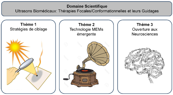
Les ultrasons (US) thérapeutiques sont en constante évolution. Pour améliorer les soins aux patients, des thérapies personnalisées - focales ou conformationnelles - doivent permettre de mieux cibler les sites pathologiques. Ces stratégies restent complexes à implémenter, car elles nécessitent un contrôle avancé : de l’émission US ; des interactions biophysiques « US/tissus ». Pour guider ces procédures, la bimodalité US (imagerie/thérapie) et l’imagerie multimodale(ex : US, IRM, modélisation, fusion) sont prometteuses. Nos recherches transversales ont été réparties selon 3 thèmes : 1)nouvelles stratégies de ciblage ; 2) technologie MEMs émergente ; 3) ouverture aux neurosciences.
Thème 1 – En chirurgie ouverte, nous avons proposé une focalisation ultrasonore - torique - pour la thérapie focale des métastases hépatiques par ultrasons focalisés de haute intensité guidés sous imagerie US. Les ablations tissulaires obtenues en préclinique étaient volumineuses, peu affectées par la vascularisation et les mouvements physiologiques (épilogue : Phases I-IIa cliniques). En radiologie interventionnelle, l'étude d’expositions ultrasonores collimatées - mono- puis bi-fréquentielles - permettait la génération d’ablations conformationnelles sous IRM (préclinique ; Phase 0 clinique), pour la thérapie transurétrale du cancer localisé de la prostate (épilogue : études multicentriques). Nous travaillons aujourd’hui sur des méthodes de visualisation temps-réel et de guidage multimodale (navigation, fusion), qui pourraient offrir de nouvelles perspectives de ciblage, mais également nécessiter une transition technologique.
Thème 2 – Pour envisager de nouvelles approches thérapeutiques avec une technologie MEMs émergente de - Transducteur Ultrasonore Capacitif Micro-usiné (CMUT) -, nous étudions la mécanotransduction (à l’échelle cellulaire) et les performances acoustiques de différents designs de réseaux d’éléments CMUTs. Outre les avantages connus (miniaturisation, multifréquence), la capacité de cette technologie à émettre des ultrasons de puissance a été démontrée. Des investigations sont en cours, avec le développement de nouvelles générations de prototypes hautement intégrés. Les thérapies ultrasonores de basse énergie pourraient aussi bénéficier de ces innovations, mais nécessitent parfois d’abord une ouverture à d’autres disciplines.
Thème 3 – Pour comprendre les mécanismes de neurostimulation par ultrasons de basse énergie, nous avons dû intégrer des techniques électrophysiologiques avancées issues des neurosciences. Dès lors, une approche comparative multi-modèle multi-échelle suggérait un rôle prépondérant de la force de radiation, dans le déclenchement des réponses neuronales. La maitrise de ces phénomènes pourrait déboucher sur de nouvelles formes de thérapies - fonctionnelles -.
Les technologies ultrasonores émergentes et les méthodes issues des computer-/neuro- sciences continueront à inspirer nos recherches, pour maitriser certains mécanismes biophysiques et proposer des approches médicales personnalisées innovantes.
Therapeutic ultrasound (US) is constantly evolving. To improve patient care, personalized therapies - focal or conformal - must allow better targeting of pathological sites. These strategies remain complex to implement, as they require advanced control: of the US emission; of “US/tissue” biophysical interactions. To guide these procedures, US bimodality(imaging / therapy) and multimodal imaging (e.g. US, MRI, modeling, fusion) are promising. Our transversal research was divided into 3 themes: 1) new targeting strategies; 2) emerging MEMs technology; 3) openness to neurosciences.
Theme 1 - In open surgery, we have proposed a - toroidal - ultrasound focusing for the focal therapy of liver metastases by high intensity focused ultrasound guided under US imaging. Tissue ablations obtained preclinically were voluminous, little affected by vascularization and physiological movements (epilogue: clinical Phases I-IIa). In interventional radiology, the study of - single then dual-frequency - collimated ultrasound exposures allowed the generation of conformal ablations under MR guidance (preclinical; clinical Phase 0), for the transurethral therapy of localized prostate cancer (epilogue: multicenter studies). We are currently working on real-time visualization and multimodal guidance methods (navigation, fusion), which could offer new targeting perspectives, but also require a technological transition.
Theme 2 - To consider new therapeutic approaches with an emerging MEMs technology of - Micro-machined Capacitive Ultrasonic Transducer (CMUT) -, we are studying the mechanotransduction (at the cellular level) and the acoustic performances of different designs of CMUT element arrays. In addition to the known advantages (miniaturization, multi-frequency), the capacity of this technology to emit high-power ultrasound has been demonstrated. Investigations are underway, with the development of new generations of highly integrated prototypes. Low-energy ultrasound therapies could also benefit from these innovations, but sometimes require opening up to other disciplines first.
Theme 3 - To understand the mechanisms of low-energy ultrasound neurostimulation, we had to integrate advanced electrophysiological techniques from neurosciences. Therefore, a multi-model multi-scale comparative approach suggested a preponderant role of the radiation force in triggering neuronal responses. The mastery of these phenomena could lead to new forms of - functional - therapies.
Emerging ultrasound technologies and methods from computer-/neuro- sciences will continue to inspire our research, to master certain biophysical mechanisms and to propose innovative personalized medical approaches.
Résumé en image (Graphical abstract)
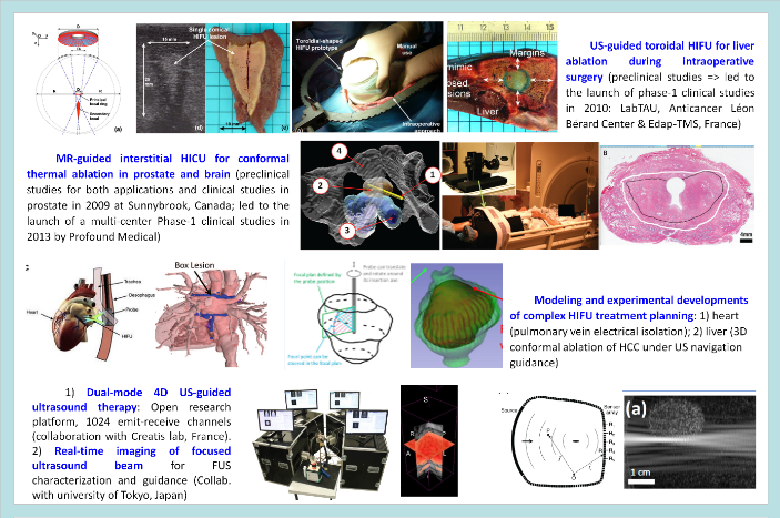
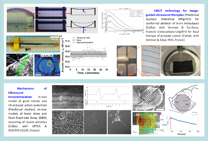
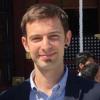
Thursday 29 April 2021
Author: Maxime Lafond
Time: 11H00
Language: French/English
Place: Conference Room at LabTAU
Abstract: The main challenge in drug delivery is crossing biological barriers like Blood-Brain Barrier, endothelium, stroma... However, there are therapeutic agents that diffuse unaffected through most barriers: bioactive gases. Bioactive gases such as Xenon (Xe) or nitric oxide (NO) can be loaded in microbubbles (MBs) to prevent their dissolution or coalescence in blood and survive passage through the lungs. MBs can be destroyed with ultrasound (US), releasing the payload that then diffuses passively. Bioactive gases find applications in cardiovascular disease. NOMBs showed bactericidal in vitro, with potential in treating infectious endocarditis. The gas payload can be released using a US imaging probe like XeMBs for neuroprotection or intravascularly with endovascular US. In all those cases, cavitation monitoring is paramount. Several methods of cavitation monitoring were investigated. The most basic way to monitor cavitation is to analyze the temporal signals to calculate the energy emitted by the MBs. This can notably help to measure a cavitation threshold and estimate the MB’s rheological properties. A combination of temporal signals from multiple sensors can provide localization information. Singular value decomposition can be implemented to separate information from cavitation, blood flow, and tissue speckle in B-mode images. Finally, frequency-domain passive cavitation imaging provides selective images of inertial and stable cavitation in a region of interest.
The video is available on our youtube channel: https://youtu.be/f0MT1UgHnwE

Thursday 22 April 2021
Author: samuel Pichardo
Time: 14H00
Language: French/English
Place: Conference Room at LabTAU
Abstract:
Dans cet exposé, nous présenterons les bases et les applications de la méthode multiaxiale dans les matériaux piézocéramiques dans la formation et la détection des ultrasons. La méthode multiaxiale consiste principalement à placer des paires d'électrodes dans une piece de matériau piézocéramique où les paires d'électrodes sont orthogonales entre elles. Pour la génération d'ultrasons, nous avons découvert qu'un champ électrique de rotation généré par un signal déphasé (mais à la même fréquence) peut contrôler la direction des ultrasons générés par la piece piézocéramique. Cette approche permettra de concevoir des sondes multi-élément où les éléments de la sonde peuvent être placés avec une distance inter-élément plus grande que les sondes traditionnelles. En mode réception, nous avons découvert que les signaux collectés par les paires d'électrodes orthogonales peuvent être combinés pour améliorer la qualité d'imagerie d’une sonde multi-élément. La méthode multiaxiale offrira des opportunités très importantes pour développer une nouvelle génération d'applicateurs et de détecteurs à ultrasons.
Video available on our youtube channel: https://youtu.be/hFuawmbiUSg
Thursday 18 March 2021
Author: Claus-Dieter Ohl
Time: 11H00
Language: English
Place: Meeting Room at LabTAU and visio Microsoft Team
Abstract: In this talk I’ll focus on the dynamics of cavitation bubbles in liquids and elastic solids mostly from experiments. In rigid gaps filled with liquids the bubble dynamics is surprisingly complex and rich. For very thin liquid gaps the elasticity of the boundaries comes into play. Then secondary cavitation can be generated from Lamb-type waves of the liquid. Recently we studied the interaction of shock waves interacting with gas bodies in a sample of gelatine. Bubble-bubble interaction and a considerable slow down of the shock front is observed.
Here is the link to the seminar:

Thursday 11 March 2021
Author: adrien Rohfritsch
Time: 11H00
Language: French
Place: Conference Room at LabTAU
Abstract:
Nous nous intéressons à la propagation d'une onde incidente à l'intérieur d'un milieu hétérogène a diffuseurs élastiques discrets. Le champ moyen, appelé onde cohérente, est caractérise par un nombre d'onde effectif complexe, qui est le cœur de notre étude. L'influence des propriétés statistiques du désordre sur ce nombre d'onde est étudiée précisément. Pour ce faire, un code numérique à hautes performances, développé au cours de ma thèse, est utilisé [1]. Cet outil permet de traiter des problèmes de plusieurs milliers de particules et prend en compte les positions réelles des diffuseurs ainsi que toutes les interactions. Ces résultats numériques sont comparés a des modèles statistiques issus de la littérature ainsi qu'à des résultats expérimentaux. Deux types de corrélation, a courte [2] et longue [3] portées, sont étudiés successivement. Ce dernier type de corrélation nous amène a considérer de nouveaux matériaux, appelés hyper uniformes [4, 5]. Des modifications très significatives des propriétés effectives sont observées qui dépendent du régime fréquentiel. Ces modifications sont en accord avec certaines théories d'homogénéisation prenant en compte la microstructure. Le travail présente ouvre la voie a la compréhension fine et a l'optimisation de matériaux fonctionnels de nouvelle génération.
[1] A. Rohfritsch, J.-M. Conoir, R. Marchiano et T. Valier-Brasier, Numerical simulation of two-dimensional multiple scattering of sound by a large number of circular cylinders, The Journal of the Acoustical Society of America, 145(6), 3320{3329, 2019.
[2] A. Rohfritsch, J.-M. Conoir, T. Valier-Brasier et R. Marchiano, Inuence of the microstructure of two dimensional random heterogeneous media on propagation of acoustic coherent waves, Phys. Rev. E, 101, 023001, 2020.
[3] A. Rohfritsch, J.-M. Conoir, T. Valier-Brasier et R. Marchiano, Impact of particle size and multiple scattering on the propagation of waves in stealthy-hyperuniform media, Phys. Rev. E, 102, 053001, 2020.
[4] O. U. Uche, F. H. Stillinger et S. Torquato, Constraints on collective density variables: Two dimensions, Phys. Rev. E, 70, 046122, 2004.
[5] Y. Jiao, T. Lau, H. Hatzikirou, M. Meyer-Hermann, J. C. Corbo et S. Torquato, Avian photoreceptor patterns represent a disordered hyperuniform solution to a multiscale packing problem, Phys. Rev. E, 89, 022721, 2014.

Thursday 14 January 2021
Author: Simon Lambert
Time: 11H00
Language: French
Place: Conference Room at LabTAU
Abstract: L’imagerie des tissus biologique en condition in vitro, ex vivo ou in vivo a connu des développements considérables ces 40 dernières années avec, en particulier, les méthodes dites translationnelles que sont le scanner X, les US et enfin l’IRM. Ces deux dernières modalités ont l’avantage d’être non invasives. La facilité de mise en œuvre des US rend cette modalité d’imagerie plus accessible, en particulier en routine clinique. Les améliorations en termes de caractérisation de la microarchitecture tissulaire avec les US proviennent essentiellement de l’accélération de la cadence d’acquisition des images. En revanche, l’IRM reste une technique difficile d’accès et assez lente comparée aux US. Néanmoins, elle permet une acquisition 3D avec une résolution spatiale pouvant, dans certaines conditions, rivaliser avec le scanner X et bien au-delà si l’on intègre une modélisation du contraste IRM qui prend naissance à l’échelle nucléaire. Le séminaire présentera les approches instrumentales et multi-échelle qui permettent de visualiser et caractériser à une échelle macroscopique, la microarchitecture tissulaire… et qui sait, peut-être même la nanostructure! L’ensemble de ces développements visent à améliorer et faciliter le suivi de l’état in vitro à l’état in vivo des tissus fabriqués en ingénierie tissulaire pour la médecine régénérative.

Wednesday 16 December 2020
Author: Cécile Fant
Time: 14H00
Language: French
Place: Visioconference (need to send a request to participate at the visio with the button below the abstact)
Abstract:
Cancer is recognized as one of today's major international health issues. Among recent advances, the rise of immunotherapies has profoundly changed the therapeutic approach. However, a sometimes partial response to treatment, the rate of recurrence or the high toxicity of certain anti-cancer agents such as radiotherapy or chemotherapy are all elements limiting the complete cure of patients. Surpassing these toxicities or improving their effectiveness are therefore crucial clinical needs. Therapeutic focused ultrasound is currently used through its thermal ablation effect to treat different types of cancer. However, its wide range of possible biological effects at the organism, tissue, cellular or molecular level, as well as its low side effects, make it a preferred modality for combination with other anti-cancer treatments to overcome their limitations. In particular, the potentiation of chemotherapy agents by focused ultrasound is an active research topic. In recent years, the study of the combination of focused ultrasound with immunotherapies has also been in full boom. In this thesis, we have studied the effect of cavitational focused ultrasound in combination with a chemotherapy agent, doxorubicin, or immunotherapies, anti-PD-1 anti-CTLA-4. A synergistic in vitro effect of the combination of ultrasound and doxorubicin was demonstrated in a mouse breast tumor model. After investigation of the underlying mechanisms, we demonstrated an additive effect of this combination on the induction of DNA double-strand breaks. This induction of DNA damage seems to occur through the release of calcium into the extracellular medium. We also studied the impact of a combination of cavitational ultrasound and immunotherapy in different in vivo mouse tumor models with partial sensitivities to different immunotherapies. After demonstrating a synergy of the combination on the control of tumor growth and the increase of overall survival in these preclinical models, an exploration of the underlying immune mechanisms was performed, highlighting the involvement of the adaptive immune system via CD8+ T lymphocytes and the increased activation of dendritic cells within the tumor environment.

Tuesday 8 December 2020
Author: Loic Daunizeau
Time: 14H00
Language: French
Place: Visioconference
Summary:
Hepatocellular carcinoma is the most common primary cancer of the liver. Interstitial thermal ablation procedures constitute a type of curative treatments for this cancer. Given the physical nature of the phenomenon used to modify temperature (radio frequency, micro wave, laser, cryotherapy), those methods may not be able to generate a conformal treatment for a given tumor shape. In some cases, this limitation may induce the thermal ablation of a large volume of non-tumor tissues. The use of an ultrasound interstitial probe mounted with a multi-element transducer capable of generating high intensity focused ultrasound (HIFU) may theoretically help to overcome this limitation. Also a transducer with an important number of elements may also provide in situ imaging. As a first step, the design of a transducer for interstitial ultrasound probe was studied. A specific configuration has been proposed for the treatment of tumors with a diameter of 4 cm. The question of the treatment planning method to adopt to reach an optimal conformal treatment has been then addressed by comparing numerical simulations of different strategies. All strategies were sufficiently conformal and none presented real assets compared to the others. Ultrasound focusing in itself provided the desired conformal thermal ablation. Finally, a robotic platform was developed for driving interstitial dual mode ultrasound probes, both in imaging and in therapy mode. This platform allowed the automatic treatment planning of in vitro tumor mimic phantoms, based on 3D ultrasound reconstruction from the B mode images obtained in situ by the interstitial probe. However, in therapy mode, the probes did not reach their specifications and did not manage to create thermal lesions in in vitro liver tissue sample. The modularity of the robotic platform allowed driving a different HIFU system, which was more robust. With this system, the platform managed to perform with success an automatic treatment planning and then the associated HIFU treatment in in vitro tissue sample.

Friday 4 December 2020
Author: Marine Sanchez
Time: 15H00
Language: French
Place: Visioconference (need to subscribe with the button below the summary)
Summary:
High Intensity Focused Ultrasound (HIFU) is a technique commonly used in the medical field for the treatment of solid tumors. This therapeutic strategy allows for the localized destruction of biological tissue by temperature elevation. This manuscript describes the design and utilization of a therapeutic system and a novel, patented focalization technique for non-invasive treatment of mammary adenocarcinomas. In a first instance, an existing device that has been previously used for intraoperative treatment of hepatic metastases, and one that is currently undergoing clinical trials, was used. An ex vivo study on human mammary samples demonstrated the possibility of applying pressures on deep tissue layers in a non-invasive manner. Based on this study, the development of a new focalization modality was developed to provide larger treated volumes while yielding treatment times under a minute and eliminating the need for mechanical steering of the device. This new modality provided treated volumes 30% larger than those produced by the existing device with no changes to treatment time. Given the limitations of the existing device, in addition to the need of integrating the new focalization strategy, a new device was designed and specifically fabricated to meet the demands for treating mammary adenocarcinomas. The natural focalization of this novel transducer allows for depositing pressure deep in tissue. When the new focalization modality is used, a larger pressure field produces increased treated volumes. The performance provided by the novel probe was first characterized through modeling studies. These results were then validated through preliminary in vitro studies on hepatic tissues as well as in ex vivo studies on human mammary tissue samples. This work represents the first stage of development of a HIFU treatment for mammary adenocarcinomas using a novel, non-invasive and image-guided HIFU device that can be used manually.
Thursday 22 October 2020
The 100% Online Scientific Game Jam took place over the weekend of 16-18 October. The organizing association Lyon Game Dev brought together young researchers and game developers to create science-themed video games. Thence, 14 games were born and the first prize was awarded to the game SonoBullewhich has been invented and developed by Maxime Fauconnier, PhD Student at the LabTAU, and his teammates from ISART Paris. SonoBulle offers an original and educational immersive playing experience for exploring the therapeutic technique of localized drug delivery by sonoporation and its physical aspects. The player controls a microbubble of gas, evolving in an environment resembling a vein, that he must approach to diseased biological cells before activating ultrasounds to permeabilize the cells and treat them. The streaming replay is still available on http://youtube.scientificgamejam.org and the game free of charge on https://kitero.itch.io/sonobulle/.
#ScientificGameJam
Monday 22 June 2020
Author: Charles-André PHILIP- Praticien Hospitalier
Service de Gynécologie-Obstetrique
CHU Lyon Croix-Rousse, 69004 LYON
Location: Conference Room - LAbTAU INSERM 1032- 151 Crs Albert Thomas 69003 Lyon;
Summary:
OBJECTIVES: In this thesis, we describe a new technique for the management of rectosigmoid endometriosis by transrectal ultrasound-guided high intensity focused ultrasound ultrasound (TR-USgHIFU). We also review several limiting factors of this technique before assessing several modifications to improve the feasibility and the efficacy of the procedure.
METHODS: We first conducted a phase I clinical trial to test the feasibility of TR-USgHIFU treatment using the FocalOne® device. We then carried out a study on anatomical specimens to assess the limiting factors of the FocaleOne® probe and to test another HIFU probe, which has smaller transducer and which is mounted on a flexible endoscope. We also performed an observational clinical study to measure the acoustic attenuation of rectosigmoid endometriosis lesions and that of normal bowel. Finally, we have developed a murine model of subcutaneous endometriosis to assess the pathophysiological effects of HIFU on endometriosis.
RESULTS: We included 23 patients in the phase I clinical trial between June 2015 and October 2019. Treatment was carried out in 20 of these patients (87% feasibility). No serious adverse events have been reported. We did not find a significant morphological effect at 6 months, but we report a significant improvement on digestive and gynecological symptoms as well as on patients’ quality of life. Studies on anatomical specimens and on MRI pictures demonstrated the role of the sacrum and the recto-sigmoid hinge in the mechanical limitations of the FocalOne® probe. The use of a smaller probe with a shorter focal could be interesting in order to increase the feasibility of the treatment. Our study on 13 patients treated surgically for rectum or sigmoid endometriosis nodule, showed that attenuation of digestive endometriosis at 3 MHz is 50.2 Np / m. This value was significantly higher than attenuation of the normal bowel (32.8; p <0.001). We finally showed that the heterologous subcutaneous murine model "BALB / c-nude # Ishikawa" was reliable and efficient to study the effects of HIFU on digestive endometriosis, as the acoustic attenuation of its nodules are remarkably close to that of endometriosis lesions. Thanks to this model, we reported that HIFU lesions are associated with a higher rate of ischemic and coagulation necrosis.
CONCLUSIONS: Treatment with TR-USgHIFU in rectosigmoid endometriosis is feasible and safe. Its morphological efficacy remains to be demonstrated, but its significant clinical efficacy is promising. It could become be a minimally invasive alternative to replace surgical treatment in this indication, especially for rectal lesions.
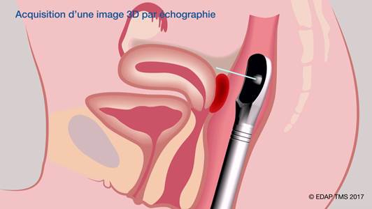
Charles-André PHILIP was born in Lyon, France, in 1986. He obtained is MD degree from University Claude Bernard (Lyon, France), in 2014 and is specialized in Obstetrics and Gynecology. He obtained his Master of Science in Oncology from Lyon 1 University (Lyon, France) during his fellowship in the Lady Davis Institute for Medical Research at McGill University (Montreal, Canada) in 2015.
His main clinical and research fields are endometriosis and oncology. He completed a 2 years fellowship in Gynecology-Obstetric in Croix-Rousse University Hospital (HCL, Lyon, France) and contributed with Pr. Gil Dubernard to the description and evaluation of 3D-Rectosonography, a new procedure combining tridimensional sonography and intra-rectal contrast for the diagnosis of rectal endometriosis

Monday 20 April 2020
Author: Marie Muller
Time: 11:00
Place: Salle de conférence de l'INSERM, 151 Cours A. Thomas
Summary: Parce qu'ils sont non-invasifs, relativement peu coûteux et faciles d’accès, les ultrasons sont une modalité d'imagerie et de caractérisation des tissus particulièrement attractive. Par ces aspects, cette modalité est plus prometteuse que toute autre pour le screening de maladies répandues (comme le cancer et l’ostéoporose, qui affectent des millions d'individus chaque année), ainsi que pour le suivi de la réponse de patients à des traitements thérapeutiques. Il y a cependant de gros inconvénients aux méthodes ultrasonores: elles manquent de spécificité, et ne sont pas adaptées à des organes a la structure complexe tels que l'os et le poumon, car la propagation ultrasonore ne s'y fait pas de manière classique. Il est toutefois possible de tirer profit de la complexité de ces milieux, pour révéler de nouvelles sources de contraste. La propagation des ondes ultrasonores dans des milieux tels que l'os et le poumon est associée à des phénomènes de diffusion qui peuvent être exploités. En modélisant la diffusion et l’atténuation ultrasonore en milieu complexe plutôt que de simplement se fonder sur des principes d’écholocation, il est possible de caractériser la microstructure de manière quantitative. Nous présenterons des exemples dans les réseaux vasculaires angiogeniques, dans l'os et dans le poumon. Nous montrerons comment ces mécanismes de diffusion peuvent être invoqués pour évaluer la malignité de tumeurs, l’ostéoporose ou des pathologies telles que la fibrose ou l'oedème pulmonaire.

Monday 17 February 2020
Author: Francesco Prada
Time: 11:00
Place: Salle de conférence 151 Cours Albert Thomas, 69003 Lyon
Summary:
Dr. Prada earned his degrees and residency at the “Università degli Studi” of Milan, Italy, perfectioning his training in major centers worldwide.
He is currently neurosurgeon in the Department of Neurosurgery at the Fondazione IRCCS Istituto Neurologico C.Besta in Milan, Italy, where he shared his interests between clinical practice, mainly focusing in skull base/pituitary surgery and neuro-oncology, and research, studying intra-operative applications of ultrasound for the treatment of cerebral and spinal tumor and vascular lesions and advanced ultrasound techniques such as Fusion Imaging for Virtual Navigation, contrast enhanced ultrasound (CEUS) and elasto-sonography.
He is now the Director of the “Ultrasound Neuro-Imaging and Therapy Lab” (UNIT-Lab), performing pre-clinical, clinical and technical research in ultrasound imaging and therapy for various neurological conditions.
He is also founder and developer of In.Tra. and Neuro-stream, start-ups related to the application of ultrasound to the brain.
Dr. Francesco Prada joined the Focused Ultrasound Foundation in Charlottesville (USA) in July 2017 where he was recipient of the Merkin Fellowship. Since July 2018 he has also been named in Brain Program Director of the Focused Ultrasound Foundation.
He is currently Visiting Professor in the Department of Neurological Surgery, University of Virginia Health Science Center in Charlottesville, Virginia (USA) where he is conducting clinical and pre-clinical researches in the field of focused ultrasound.

Thursday 13 February 2020
Author: Xiang Ji
Time: 11:00
Place: Salle de conference 15 cours Albert Thomas, 69003 LYON
Summary: Uterine fibroids are one of the most popular indications for HIFU, and over 10,000 patients are treated by HIFU all over the world every year. There are several commercial ultrasound-guided HIFU systems for treatment of uterine fibroids in China, but HIFU phased array are still rarely used in their systems. In order to take advantage of phased arrays, we have developed a B-mode ultrasound-guided HIFU phased-array system, where the guidance probe is mounted in a rotatable platform and harnessed to track the focus steered in three-dimensional volume. A three-center 120-patient clinical trial on the safety and efficacy of treatment of uterine fibroids using the developed system began in the end of 2018. Follow-ups include 3-month ultrasound imaging and 1-, 6-month magnetic resonance (MR) imaging after treatment. Currently one center has completed its treatments of patients, and the preliminary results of the non-perfused volume ratio and fibroid shrinkage as well as patient response have verified the safety and efficacy of the developed system.

Friday 20 December 2019
Author: Nicolas Asquier
Time: 10:00
Place: Rockefeller Theses Room
Summary:
The blood-brain barrier (BBB) is a natural protection of the central nervous system. However, it limits the delivery of many drugs to the brain tissues. It can be temporarily disrupted by ultrasound exposure combined with intravenous injection of microbubbles. In this manuscript, BBB disruption with an implantable unfocused ultrasound device is studied.
An automatic method for quantifying the volume of BBB disruption using MR images from a phase 1/2a clinical study in patients with reccurent glioblastoma was assessed and validated. A correlation between the probability of disruption and the local acoustic pressure was found.
Microbubbles cavitation activity was studied in vitro to better understand its effect on BBB disruption. The uncertainty on the amplitudes of cavitation signals recorded with a passive single-element detector (PCD) through the skull was quantified. A position-based correction of the PCD signal was assessed and validated.
The effect of the volume of a cavitation cloud in the unfocused ultrasound field on the signal amplitude recorded by the PCD during the clinical treatment was discussed. Two methods for localizing and discriminating cavitation sources in a transcranial context were evaluated by simulations and in vitro.
Thursday 19 December 2019
Author: Victor Barrère
Time: 13:30
Place: Salle des thèses de Rockefeller
Summary:
Les Ultrasons Focalisés de Haute Intensité (HIFU) sont une technique de traitement non invasive par effet thermique largement utilisée en clinique. Les interventions peuvent être monitorées par Imagerie par résonnance Magnétique (IRM) ou par échographie. L’IRM permet la réalisation d’excellentes images anatomiques ainsi qu’une visualisation fiable des nécroses et une mesure de l’augmentation de température générée lors des interventions. Elle présente cependant des problématiques de coût, de disponibilité et de compatibilité avec les patients et les dispositifs. L’échographie est beaucoup plus accessible et moins onéreuse, avec des images de haute résolution spatiale et temporelle, mais ne permet pas le suivi de la température. L’objectif de ce travail est d’évaluer si les informations contenues dans le signal rétrodiffusé par les tissus au cours d’un traitement peuvent être utilisées pour réaliser un monitoring de la création des lésions HIFU.
Les expériences menées ont permis d’investiguer la possibilité de réaliser un monitoring ultrasonore de la température pendant les interventions. En particulier, l’énergie du signal rétrodiffusé a pu être évaluée. L’augmentation linéaire de ce paramètre avec la température au cours du chauffage ont permis la réalisation de cartes de température en 2D pendant l’intervention.
La plupart des paramètres acoustiques connaissent une évolution non linéaire en fonction de la température, notamment au-delà de 50°C, température à partir de laquelle apparaissent des phénomènes de coagulation et de restructuration cellulaire. Des analyses par ultrasons quantitatifs (QUS) couplées à une analyse histologique ont permis d’étudier la transformation de la microstructure du tissu dans des tissus exposés à des températures allant de 37°C à 80°C, en vue d’expliquer les changements de valeur des paramètres ultrasonores au cours du traitement. La taille moyenne des diffuseurs et leur concentration acoustique moyenne ont pu être évaluées, ainsi que la taille moyenne des cellules et l’espace intercellulaire.
Le monitoring des interventions a pu également être investigué par élastographie passive. La vitesse de cisaillement moyenne a été évaluée au cours d’un chauffage ultrasonore dans la région de la lésion, et des cartes d’élasticités ont été calculées pendant le chauffage pour réaliser des films de la croissance des zones traitées.
Enfin les techniques de caractérisations développées au laboratoire ont pu être exportées au bloc chirurgical pour la caractérisation ultrasonore de pièces d’hépatectomie ex vivo contenant des tumeurs primaires et secondaires.

Monday 25 November 2019
Author: Dr Mario Ries – Image Sciences Institute-University Medical Center Ultrecht
Time: 2 PM
Place: LabTAU – Conference Room
Summary:
While therapeutic HIFU applications in the linear pressure regime have meanwhile established their place in numerous clinical applications, non-linear acoustic phenomena have despite their considerable potential so far been left largely unexploited.
Here we contrast the in-vivo experience of the UMC-Utrecht with non-linear acoustic applications for local control vs systemic therapy. Shock wave enhanced HIFU thermotherapy can hereby overcome the typical limitations of HIFU ablations in high perfused organs such as the liver, while mechanical tissue fractionation allows transforming immunologically 'cold' Neuroblastoma tumors into responsive 'hot' tumors, which in combination with immune checkpoint inhibitors lead to a systemic response.

Thursday 21 November 2019
Author: Zhishen Sun
Summary: The Lorentz force, as the linkage between the mechanical displacement and the electrical current, is investigated in this thesis in two fields of the medical acoustics, i.e. scanning electric conductivity gradients with ultrasonically induced Lorentz force (SECG-UILF) and elastography.
The first part of the thesis studies the SECG-UILF. To reduce the instantaneous stimulation power to the transmitting transducer and at the same time the peak acoustic pressure from the transducer, this thesis proposes to apply the linearly frequency-modulated ultrasound pulse excitation or the sinusoidal step-frequency ultrasound pulse excitation in SECG-UILF. The second part of the thesis studies elastography which uses the cross-correlation approach of diffuse field to reconstruct the shear wave velocity. First, generation of shear wave sources on the soft medium surface is investigated by stimulating a non-ferromagnetic conductive ring or patch with a transient magnetic field. Due to the Faraday's law of induction, an eddy current is generated remotely in the conductive ring or patch by the transient magnetic field. The eddy current at the same time couples with the magnetic field, generating the Lorentz force, which constitutes a torque on the ring or patch and vibrates the ring or patch. The origin and the frequency and amplitude characteristics of the Lorentz force acting on the conductive ring are confirmed by the displacement measurement using an interferometric laser probe: (1) the eddy current induced by the transient magnetic field and the orthogonal component of the magnetic field constitute the two essential elements of the Lorentz force; (2) the carrier frequency of the Lorentz force equals the sum of the carrier frequencies of the stimulating transient magnetic field and its time derivative; and (3) the amplitude of the Lorentz force increases quadratically upon the amplitude of the stimulating current to the coil. Under a transient magnetic field of changing rate of 10.44 kTs-1, the patch generates a shear wave field source of amplitude of 100 μm at the surface of the sample of polyvinyl alcohol (PVA) phantom. The shear wave fields created and propagating in the PVA phantom by experiments agree qualitatively well with the theoretical shear wave fields calculated through the analytical Green function solution. The potential of the generated shear wave fields for the cross-correlation based shear wave velocity reconstruction is explored. The displacement fields in the multi-shaped soft biological tissue mimicking agar phantoms are recorded at a sampling rate of 1000 frames/s as the multiple shear wave sources propagate in the phantoms. Based on the cross-correlation approach, the shear wave velocity maps are reconstructed from 100 frames of the displacement fields, which show that: (1) the interfaces or boundaries between regions of different stiffness can be clearly recognized, which are completely concealed in the ultrasound images; (2) cylindrical inclusions of diameter as small as 5 mm can be differentiated from the background, but at the same time, there are phony boundaries in the homogeneous areas which undermine the performance of the method; and (3) using the cross-correlation approach, it is possible to qualitatively but not quantitatively reconstruct the shear wave velocity in the samples from 100 frames of the displacement fields.
Time:13h30
Place: Salle de réunion du LabTAU, 151 Cours Albert Thomas, Lyon

Tuesday 19 November 2019
Author: Claude INSERRA
Summary: Contrôle de la dynamique spatiotemporelle de bulles de cavitation ultrasonore -
Une bulle acoustique est un oscillateur non linéaire, excité par un champ ultrasonore, qui possède un grand nombre d'avantages pour certaines applications thérapeutiques, en tant que vecteur localisé de l'énergie acoustique, avec une forte échogénicité, et aidant au transport de médicament au sein de cellules ou tissus biologiques. Les localiser in vivo, et détecter/quantifier leur activité de cavitation est primordial pour assurer une meilleure efficacité des traitements ultrasonores. Afin de les détecter de manière extra-corporelle, des signatures acoustiques propres à la bulle sont classiquement utilisées. Ces signatures sont (i) le bruit large bande, caractéristique de l'implosion de bulles, qui identifie le régime de cavitation inertielle et (ii) la composante sous-harmonique (entre autres composantes) de la fréquence fondamentale, dont les origines sont diverses, mais qui identifie un régime temporellement stable d'oscillations (ou d'implosions périodiques d’un nuage de bulles). A l'aide d'une écoute passive de ces signatures, on peut distinguer l'activité de cavitation selon ces deux régimes.
Dans une première partie de ce mémoire le contrôle temps-réel de l'activité de cavitation d'un nuage de bulles est présenté. Ce contrôle permet de s'affranchir du caractère stochastique de la cavitation ultrasonore, et d'assurer un niveau de cavitation reproductible, temporellement stable. Deux stratégies de contrôle sont présentées, une sur l'indice de cavitation inertielle, l'autre sur l'indice de cavitation stable. Elles reposent sur la modulation en temps-réel de la tension électrique appliquée aux transducteurs ultrasonores. Cette modulation est régie par un processus proportionnel de contrôle afin d'assurer un niveau de cavitation donné par l'opérateur. Grâce à une programmation sous FPGA (Field Programmable Gate Array), des temps de rétroaction inférieurs à la milliseconde ont permis le contrôle de cavitation au sein de tirs ultrasonores pulsés. Il est montré que la stratégie de contrôle permet de s'affranchir de l'utilisation d'agents de contraste ultrasonore, au moins in vitro. Les taux de sonoporation/transfection obtenus lors de l'utilisation du contrôle sont généralement plus reproductibles que ceux obtenus en séquences ultrasonores à intensité acoustique fixe. L'utilisation du contrôle de cavitation stable a mis en évidence la possibilité de sonoporer des cellules tout en minimisant la mortalité cellulaire. Afin de saisir l'origine physique de la sonoporation cellulaire (en termes d'interaction bulle-cellule), il est nécessaire de différencier les régimes de cavitation stable et inertiel dans les phénomènes cellulaires étudiés.
Dans une seconde partie de ce mémoire mes travaux sur la dynamique temporelle de bulle unique sont présentés. Après des balbutiements sur la dynamique de bulles présentant des couplages translation/oscillation radiale en champ acoustique intense, une étude analytique sur la dynamique de bulle en excitation bifréquentielle est réalisée. Cette étude permet de démontrer l'intérêt de la génération de nouvelles composantes fréquentielles proches de la fréquence de résonance de la bulle, qui se décale par effet non linéaire. Une démarche expérimentale a été initiée pour vérifier ces travaux théoriques avec la conception d'une chambre de lévitation acoustique où une bulle unique est piégée sur un ventre de pression du champ ultrasonore. L'intérêt de cette configuration est d'obtenir les oscillations d'une bulle loin de toute paroi, et de faciliter les observations par caméra rapide. En utilisant un champ acoustique modulé en amplitude, une déviation des oscillations radiales de la bulle est observée. L'interface se déforme selon des modes de surface qui sont périodiquement déclenchés, maintenus puis annihilés. Ce déclenchement périodique des modes de surface de bulle a mis en évidence le couplage non linéaire existant entre les modes de volume, de translation et de surface de la bulle. L'ensemble des observations expérimentales a ainsi permis de valider les plus récents modèles théoriques prenant en compte ces couplages. Les oscillations non sphériques de bulle ont finalement été maîtrisées par utilisation d'une technique de coalescence entre deux bulles filles. Cette technique a permis le contrôle de l'orientation des déformations non sphériques, ainsi que la mise en place d'un régime permanent des modes de surface. Profitant de ce régime permanent, l'ensemencement de particules dans la chambre de lévitation acoustique permet l'observation des écoulements fluides induits par les oscillations de bulles. Les motifs d'écoulements observés sont reliés à la dynamique de la bulle, mais aussi à la position de la bulle dans la zone d'instabilité des modes de surface. Des motifs confinés (proche de l'interface de la bulle) ou en croix (très étendu jusqu'à plusieurs diamètres de bulle) sont relevés. Pour expliquer ces motifs, un modèle analytique des écoulements induits par une bulle axisymétrique oscillant sur ses modes de volume, translation et de surface, est réalisé. Il permet le calcul exact du champ de vitesse moyen de l'écoulement induit. Une classification des motifs expérimentaux à l'aide de ce modèle analytique est en cours.
Time:13:30
Place:Salle de Conférence de l'INSERM, 151 Cours Albert Thomas, LYON
Thursday 14 November 2019
Author: Maxime Polichetti
Summary:
Ce travail s'intéresse au suivi spatio-temporel par imagerie ultrasonore de la cavitation acoustique, utilisée au cours de certaines techniques de thérapie par ultrasons, correspondant à la formation de bulles de gaz qui oscillent et éclatent.
Initialement, la méthode TD-PAM (Time Domain Passive Acoustic Mapping, en anglais), a été développée pour cartographier l’activité de cavitation à partir des signaux acoustiques émis par les bulles, enregistrés passivement par une sonde linéaire d'imagerie ultrasonore. Toutefois, le TD-PAM souffre d’une trop faible résolution et de nombreux artefacts de reconstruction. De plus, il est lourd en temps de calcul car il est formalisé dans le domaine temporel (TD).
Pour pallier ces deux limitations ce travail propose des méthodes avancées d'imagerie ultrasonore passive de la cavitation. La présentation s'articule autour de deux contributions principales :
• La notion de matrice de densité inter-spectrale a été introduite pour l'imagerie de la cavitation. Dès lors, quatre méthodes d’imagerie dans le domaine de Fourier (FD) ont été étudiées, adaptées et comparées : le FD-PAM (non-adaptatif), la méthode Robuste de Capon FD-RCB (adaptatif, par optimisation), le Functional Beamforming FD-FB (adaptatif, par compression non-linéaire) et la méthode MUltiple Signal Classification FD-MUSIC (adaptatif, par projection en sous-espaces).
• Les performances de ces méthodes FD ont été étudiées expérimentalement in vitro cuve d’eau avec une comparaison par imagerie optique. Les méthodes adaptatives FD proposées ont démontré leur potentiel à améliorer le suivi spatio-temporel des bulles. Le FD-RCB offre une localisation supérieure au FD-PAM mais souffre d'une importante complexité algorithmique. Les performances du FD-FB sont intermédiaires à celles du FD-PAM et du FD-RCB, pour une complexité de calcul équivalente au FD-PAM. Le FD-MUSIC a le potentiel de mettre en évidence de faibles sources acoustiques, mais ne conserve pas leurs quantifications relatives
Time:11:00
Place: Salle de conférence, INSERM 151 Cours A.Thomas, LYON

Wednesday 6 November 2019
Author: Francis Bessiere
Summary:
A la croisée des chemins entre médecine et physique des ultrasons, ce travail de thèse s’est intéressé à l’apport de solutions diagnostiques et de thérapeutiques novatrices dans le domaine de l’électrophysiologie cardiaque.
Un système capable de délivrer des ultrasons focalisés dans le cœur par voie transoesophagienne sous guidage par ultrasons a été développé et testé in vivo chez 6 porcs. Les tirs HIFU ont été délivrés sur les oreillettes et les ventricules. Lors de l'autopsie, une analyse visuelle a démontré la présence de lésions thermiques dans les zones ciblées chez 3 animaux. Ces lésions ont été confirmées par analyse histologique (taille moyenne: 5,5 mm2 x 11 mm2). Aucune lésion thermique œsophagienne n'a été observée. Un animal a présenté une bradycardie due à un bloc auriculo-ventriculaire, ce qui a permis de confirmer une réelle interaction entre les tirs HIFU et le tissu nodal cardiaque. Nous avons cependant observé un manque de précision, principalement lié aux mouvements cardiaques ainsi qu’aux structures anatomiques situées entre les zones ciblées et le transducteur de thérapie. Ces difficultés ont été principalement reliées à l’anatomie du modèle porcin, loin de celle de l’être humain. La recherche d'un meilleur modèle a conduit à des tests d'imagerie concluants sur des babouins.
Des expériences supplémentaires ont été conduites afin d'améliorer la cartographie des arythmies ventriculaires et le suivi de la formation de lésions pendant l'ablation.
Des expériences ont été menées sur les ventricules gauches de quatre coeurs de porcs en mode travaillant. Le protocole visait à démontrer que différents modèles d'activation mécanique pouvaient être observés, que le ventricule soit en rythme sinusal, stimulé depuis l'épicarde ou l'endocarde. Des acquisitions d’imagerie de déformation électromécanique (EWI) ont été enregistrées sur les faces antérieures, latérales et postérieures du ventricule gauche. Elles ont été ensuite analysées à l’aveugle par deux lecteurs indépendants.
Les interprétations des séquences EWI étaient correctes dans 89% des cas. Le taux de concordance globale entre les deux lecteurs était de 83%. Dans un ventricule stimulé, l'origine du front d'onde était focale et provenait de l'endocarde ou de l'épicarde stimulé. En rythme sinusal, le front d'onde était activé depuis tout l'endocarde, en direction de l'épicarde, à une vitesse de 1,7 ± 0,28 m.s-1. Les vitesses du front d'onde ont été mesurées respectivement lorsque l'endocarde ou l'épicarde étaient stimulés à une vitesse de 1,1 ± 0,35 m.s -1 et 1,3 ± 0,34 m.s-1 (p = NS).
Nous avons aussi démontré sur des échantillons ex-vivo que l'imagerie trans oesophagienne par analyse des ondes de cisaillement (élastographie) pouvait cartographier l'étendue des lésions HIFU.
Des tirs HIFU ont été réalisés à l'aide de la sonde trans oesophagienne sur des échantillons de blanc de poulet (n = 3), puis sur un modèle porcin ex vivo d'oreillette (gauche, n = 2) et de ventricule gauche (n = 1). L’élastographie a fourni des cartes de rigidité des tissus avant et après l'ablation. Les zones des lésions ont été obtenues par analyse et quantification des changements de couleur des tissus puis ont été comparées aux images par élastographie.
Dans le blanc de poulet, la rigidité est passée en moyenne de 4.8±1.1 kPa à 20.5±10.0 kPa (ratio 5.0±3.2). Dans le ventricule gauche, la rigidité est passée en moyenne de 21.2±3.3kPa à 73.8±13.9kPa (ratio 3.7±1.2). Dans l’oreillette gauche, la rigidité est passée en moyenne de 12.2±4.3 kPa à 30.3±10.3 (ratio 3.2±2.0). En histologie, la taille des lésions variait de 0.1 à 1.5 cm2 dans la zone du plan d'imagerie. Les caractéristiques morphométriques étaient similaires entre histologie et élastographie.
Time:14:00
Place:Salle de conférence de l''INSERM, 151 Cours Albert Thomas, LYON

Monday 14 October 2019
Author: Johannes Aichele
Summary:
La première partie se penche sur le comportement des ondes dans des milieux poreux. La dispersion de l'onde de cisaillement et de l'onde de compression secondaire dans des matériaux imitant le poumon est analysées expérimentalement, puis comparées à la théorie de poro-élasticité de Biot. Les résultats quant à l'onde de cisaillement sont conformes à la théorie, et ceux de l’onde de compression y correspondent qualitativement. Pour conclure, dans le cas des milieux mous, poreux et élastiques, la dispersion des ondes élastiques est gouvernée par les propriétés du fluide visqueux. Ainsi, les résultats présentés au cours de cette thèse pourraient consolider la théorie nécessaire à une imagerie de l’élasticité pulmonaire. Potentiellement d’autres organes pourraient en bénéficier : en effet, le foie et la rate sont constitués d'un pourcentage élevé de sang. Ce fluide non-Newtonien présente une forte variabilité de la viscosité. Celle-ci implique la nécessité d’étudier le rôle du sang vis-à-vis de l'élastographie du foie, pas encore expliquée par les modèles visco-élastique. En outre, la preuve expérimentale de l'onde de compression secondaire est utile dan l’étude de la propagation d'ondes poro-élastiques. Il est important de noter que l’onde de compression secondaire a été objet principal d’études approfondies en géophysique et n’a été observée que dans quelques rares cas. La partie suivante s’intéresse à une problématique de la géophysique explorée au moyen de l’imagerie de l’onde de cisaillement :Que pourrait révéler sur la nature de la rupture dynamique d’une instabilité de friction l’étude du champs d’onde élastique ? Comment la rupture, le processus responsable des tremblements de terre, est générée? En observant la carte du champ d’onde de cisaillement pendant la rupture d’une aspérité granulaire au point source et dans le milieu, il est possible d’acquérir une meilleure compréhension de la propagation de la rupture. L’expérience qui se base sur l’utilisation de fantômes mous et élastiques montre une multitude de phénomènes qui sont aussi présents dans la friction des roches en laboratoire, ainsi que dans la Terre. Par exemple, les ruptures supershear, sub-Rayleigh, une phase de nucléation et la friction du type stick-slip ont été observés. Toutefois, ni un mécanisme de source d’une force unique, ni un double-couple n’arrivent à expliquer l’ensemble des ruptures observées. De plus, pour analyser la complexité spatio-temporelle des ruptures de cette expérience, un workflow semi-automatique intègrant la segmentation d’image et la vision numérique est suggéré.
Time:13:30
Place:Salle de Séminaire
Friday 4 October 2019
Author: Sarah Clève
Summary: Microbubbles find use in several domains, one of them being medical ultrasound applications. Different characteristics of those bubbles such as their acoustic resonance or their destructive effect during inertial cavitation can be exploited. Another phenomenon induced around acoustically excited bubbles is microstreaming, that means a relatively slow mean flow with respect to the fast bubble oscillations. Microstreaming and its associated shear stresses are commonly agreed to play a role in the permeabilization of cell membranes, a detailed understanding of the induced flows is however missing. To acquire basic physical knowledge, this work focuses on the characterization of streaming induced around an air bubble in water, more precisely around
a single acoustically trapped and excited, nonspherically oscillating bubble. The experimental part consists of two steps. First, the bubble dynamics, in particular the triggered shape mode and the orientation of the bubble have to be controlled. For this, the use of bubble coalescence proves to be an adequate method. In a second step, the microstreaming is recorded in parallel to bubble dynamics. This allows to correlate the obtained streaming patterns to the respective shape oscillations. The large number of obtained pattern types can be classified, in particular with respect to the mode number and bubble size. A close investigation of the bubble dynamics allows furthermore deducing the important physical mechanisms which lead to such a variety of streaming patterns. In order to confirm the experimental findings, an analytical model has been developed. It is based upon time-averaged second-order fluid mechanics equations and the experimentally obtained bubble dynamics serves as input parameters. Supplementary to the microstreaming work, this manuscript contains a short section on directed jetting of contrast agent microbubbles, which might appear at high acoustic driving. The impact of those microjets on cell membranes presents another mechanism made responsible for the permeabilization of cell membranes.
Time: 13h30
Place: Ecole Centrale Lyon - Bâtiment W1 Amphi201

Thursday 27 June 2019
Author: Xueding Wang
Summary:
Photoacoustic imaging (PAI), also referred to as optoacoustic imaging, is an emerging biomedical imaging technology that is noninvasive, nonionizing, with high sensitivity, satisfactory imaging depth and good temporal and spatial resolution. Like conventional optical imaging, PAI presents the optical contrast which is highly sensitive to molecular conformation and biochemical contents of tissues and can aid in describing tissue metabolic and hemodynamic changes. Unlike conventional optical imaging, the spatial resolution of PAI is not limited by the strong light diffusion but instead determined by the measurement of light-generated ultrasonic signals. As a result, the resolution of PAI is parallel to high-frequency ultrasonography.
At the University of Michigan School of Medicine, our research has been focused on clinical applications of PAI, including arthritis, cancer, liver conditions, Crohn’s disease, and eye diseases. In this talk, I will introduce some of our recently development of PAI technologies, including 1) development of point-of-care PAI system for human inflammatory arthritis, and 2) development of quantitative PAI for evaluating histological microfeatures and microenvironment of cancer. I will also present our recent development of a photoacoustic based anti-vascular technology named photo-mediated ultrasound therapy (PUT). Using a combination of a low intensity laser concurrently with ultrasound, PUT can noninvasively remove microvessels without damaging surrounding biological tissue, and shows potential to the treatment of a variety of diseases associated with neoangiogenesis, such as age-related macular degeneration and diabetic retinopathy.
Time:15:00
Place:Salle de conférence
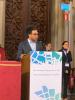
Tuesday 18 June 2019
Dr. W. Apoutou N’DJIN, Ph.D., Research Associate in Biomedical Engineering & Physics at the Laboratory of Therapeutic Applications of Ultrasound (LabTAU, French National Institute of Health and Medical Research, University of Lyon, France), was recently presented the International Society of Therapeutic Ultrasound’s (ISTU) Frederic LIZZI Early Career Award at the 19th International Symposium for Therapeutic Ultrasound. One of ISTU’s most prestigious awards, the Frederic LIZZI Early Career Award was established in 2008 to honor Dr. Frederic L. LIZZI, a pioneer in advanced diagnostic and therapeutic applications of ultrasound. Since then, it is presented to one recipient annually (except in special circumstances) for their contributions to therapeutic ultrasound. The recipient should be within 10 years of their terminal degree and is selected among a pool of nominees by ISTU’s Organizing Committee and Board.
The event was held in Barcelona (Spain) at the occasion of the first joint meeting of the ISTU Society and the European Focused Ultrasound Charitable Society (EUFUS). The Symposium has been held at the historic building of the Universitat de Barcelona, one of the most emblematic sites in the city centre, which was constructed in 1863 by the architect Elies Rogent.
This year’s LIZZI Award was shared by two researchers, Dr. W. Apoutou N’DJIN, Ph.D. (LabTAU, INSERM, CLB, Univ Lyon, France) and Dr. Kevin J. HAWORTH, Ph.D. (College of Medicine, University of Cincinnati, United States). Following ISTU tradition, LIZZI award recipients delivered the LIZZI Award Lecture and shared a dedicated session with the 2019 FRY Award (Pr. Chrit MOONEN, Ph.D., Division of Imaging, Uninversity Mecial Center Utrecht, Netherland) and the 2019 PIONEER Award (Pr. Leonid GAVRILOV, Ph.D., Andreyev Acoustics Institute, Physical Acoustics, University of Moscow, Russia).
W. Apoutou N’DJIN delivered a LIZZI Lecture entitled " A healthy future for focal conformal therapeutic ultrasound" and highlighted some of their contributions in the development of new ultrasound targeting strategies and technologies in surgery and interventional radiology, as well as their openness to methods coming from neurosciences for studying ultrasound neurostimulation.

Thursday 28 March 2019
Author: Nicolas Benech
Summary: In this work, we investigate, the feasibility of quantifying the Young's modulus of breast tissue from the auto-correlation of a diffuse acoustic field computed from a sequence of B-mode images acquired through conventional ultrasound scanners. Inspired in the seismological approach of retrieving the Green's function by cross-correlation of diffuse fields, we obtained a quantitative expression that relates the local shear modulus of soft tissue with the auto-correlation of the displacement field generated by the presence of an acoustic diffuse field in the medium. In addition, we designed a mechanical prototype device adaptable to the breast anatomy, in order to create the necessary conditions in terms of diffuse field generation. The displacement field was measured from a sequence of B-mode images acquired at standard frame rates (30 – 50 Hz) with standard ultrasound scanners (Toshiba Nemio NX and SIUI Apogee 3800). This method was tested in a breast tissue mimicking phantom. We also performed an in-vivo measurement as a preliminary validation. In this last case, the reconstructed images are in accordance with the patient known pathology (BI-RADS 5, Infiltrating Ductal Carcinoma, Score 6). The pathological breast shows a heterogeneous elasticity map with a mean Young’s modulus of 98.1 ± 6.3 KPa, while the normal breast presents a homogeneous map with a mean Young’s modulus of 24.7 ± 8.1 KPa.
Time:11:00
Place:Salle de conférence de l'INSERM, LabTAU u1032 151 Cours Abert Thomas, LYON 03
Wednesday 27 March 2019
Author: Jeremy Vion
Summary: Focused ultrasound (US) are considered as a promising tool to develop a new modality of therapeutic neurostimulation. An in vivo invertebrate nervous model (system of giant fibers of the common earthworm, Lumbricus terrestris) has been proposed to study the biophysical mechanisms underlying the phenomenon of US neurostimulation. After proving the feasibility, the nervous responses associated with different stimulation modalities (mechanical, electrical, US) were characterized and compared together. Following a causal approach of interpretation, the afferent nerves were deduced to be the locus of interaction between the nervous system and the US stimulus (pulsed, 1.1 MHz, 2% DT, 2.5 kW/cm²). The respective influences of each acoustic parameter on the neurostimulation success rate were evaluated. Results suggest afferent nerves are sensitive to the value of the ‘mean radiation force’ carried by the US stimulus, whatever combination of parameters leading to it. Complementarily, the role played by cavitation in the phenomenon of US neurostimulation was investigated, using both the monitoring of cavitation indexes and ultra-fast imaging. It was concluded that neither the occurrence of stable cavitation nor particularly high levels of inertial cavitation were necessary conditions for the occurrence of US-induced nervous responses. A new experimental campaign was launched, aiming to apply the methodology developed for the invertebrate nervous model to a nervous model of higher complexity. The feasibility of using Microelectrode Arrays (MEA) to record US-induced nervous responses from mouse cortical slices was demonstrated, and further trials are currently undertaken.
Time: 13h
Place: LabTAU Conference Room
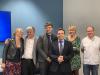
Tuesday 19 March 2019
Author: Laurent Milot
Summary: Les métastases hépatiques du cancer colorectal (MHCR) sont fréquentes et sont associées à une mortalité significative. Ces dernières décennies, des progrès thérapeutiques importants ont permis d’en améliorer le pronostic. Plus particulièrement, le rôle des résections hépatiques s’est considérablement élargi dans la maladie métastatique limitée au foie, conduisant à un changement radical dans la prise en charge. Ceci a naturellement eu des répercussions sur l’imagerie, qui doit être très performante au niveau lésionnel, nécessitant des sensibilité et spécificité très élevées. Si les techniques modernes ont permis une amélioration très nette en termes de sensibilité, en particulier grâce aux produits de contraste hépatospécifiques et de l’imagerie pondérée en diffusion, l’amélioration de la spécificité est moins claire et moins bien évaluée. Pourtant, la spécificité est tout aussi importante dans ce contexte, où les erreurs diagnostiques sont coûteuses, avec des chirurgies inutiles en cas de faux positifs, et des résections incomplètes en cas de faux négatifs. Ces deux situations sont accompagnées d’une morbi- mortalité très importante. Le présent travail de thèse va donc explorer de nouvelles pistes dont l’objectif ultime serait d’améliorer la spécificité de l’imagerie des MHCR.
La première étude confronte l’apparence des métastases d’origine colorectale en IRM de haute résolution et leur histologie sous-jacente. Cette étude originale démontre que la fibrose tumorale apparait en hypersignal T2 et la nécrose tumorale en hyposignal T2 et hypersignal T1, ce qui va à l’encontre du dogme classique. La seconde étude explore la faisabilité de la fusion d’images IRM/échographie dans l’exploration de lésions hépatiques focales chez des patients ayant un cancer colorectal. Cette étude montre qu’un nombre significatif de lésions ne peuvent être visualisées à l’échographie qu’en utilisant la fusion, ouvrant la voie à une meilleure caractérisation lésionnelle en combinant les atouts de l’échographie et de l’IRM. Enfin, la troisième étude, complétée d’une revue iconographique, analyse le comportement IRM des lésions hépatiques après injection d’un produit de contraste intravasculaire. Elle montre une accumulation progressive du contraste au sein des angiomes, mais pas dans les métastases, conduisant à des apparences très différentes sur la phase tardive. Ceci était aussi observé dans les lésions de petites tailles, ce qui devrait permettre une meilleure spécificité dans les cas difficiles.
Time: 11h
Place: Hôpital Edouard Herriot Pavillon B Grande Salle de réunion
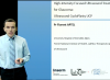
Monday 4 March 2019
Dear all
The 12th course composing the MOOC is now available. This clinical presentation is given by Professor Florent APTEL on «HIFU for treating glaucoma»
Note that this is the last of the 12 courses composing this first MOOC dedicated to therapeutic ultrasound but that all courses remain available online
Note that this is the last of this first MOOC dedicated to therapeutic ultrasound and that all the courses will still be available on-line after March 4.
You can register on Claroline by clicking here
We hope you enjoyed the presentations and again we wanted to thank you for showing your interest in the first MOOC dedicated to therapeutic ultrasound.
If you have any suggestion for further Courses on therapeutic ultrasound please let us know!
We are looking forward to your feedback.
Thank you
Monday 25 February 2019
Author: Gilles THOMAS
Summary: Kidney Stones can be found in the kidney, ureter, or in the bladder, and affect about 1 in 11 people at least once in a lifetime in the US. Extracorporeal shock wave lithotripsy is a widely used technique where high intensity acoustic pulses are focused toward kidney stones in order to break them. The work presented in this thesis focus on optimizing piezoelectric lithotripter, both in design and efficiency, in order to have more efficient treatment while also being less costly. First, a study of the current state of lithotripsy was made, followed by experiments on commercial and experimental lithotripters in order to define properly the different parameters to be worked on. From this, it was decided to optimize the current piezoelectric elements in the lithotripter to obtain a more efficient treatment. Then, a lithotripter using optimized lens focused piezoelectric transducers set in confocal setups was designed and manufactured. Its acoustic characterization, effect on cavitation and model stone fragmentation efficiency were evaluated. The resulting lithotripter showed performances equivalent to existing commercial lithotripter, while allowing more flexible treatment than traditional lithotripter.
Time: 14H
Place: Conference Room, LabTAU, 151 Cours Albert Thomas, 69003 LYON

Tuesday 19 February 2019
Dear all
The 11th course composing the MOOC is now available. This clinical presentation is given by Professor Franco ORSI on «Application of HIFU in oncology »
It will be followed on March 4 by a course given by Professor Florent APTEL on «HIFU for treating glaucoma».
You can register on Claroline by clicking here
Hope you’ll enjoy the presentation!
We are looking forward to your feedback.
Thank you for showing your interest in the first MOOC dedicated to therapeutic ultrasound
Tuesday 5 February 2019
Dear all
The 10th course composing the MOOC is now available. This clinical presentation is given by Professor Pejman GHANOUNI on «MR guided high intensity focused ultrasound» : A pediatric application».
It will be followed on February 18 by a course given by a Professor Franco ORSI on «Application of HIFU in oncology».
You can register on Claroline by clicking here
Hope you’ll enjoy the presentation!
We are looking forward to your feedback.
Thank you for showing your interest in the first MOOC dedicated to therapeutic ultrasound
Monday 21 January 2019
Dear all
The 9th course composing the MOOC is now available. This technical presentation is given by Professor Bruno QUESSON on «MRI for guiding HIFU treatment».
It will be followed on February 4 by a course given by Professor Pejman GHANOUNI on «MR guided high intensity focused ultrasound : A pediatric application»
You can register on Claroline by clicking here
Hope you’ll enjoy the presentation!
We are looking forward to your feedback.
Thank you for showing your interest in the first MOOC dedicated to therapeutic ultrasound
Tuesday 8 January 2019
Dear all
The 8th course composing the MOOC is now available. This clinical presentation is given by Professor A. CARPENTIER on «Opening the BBB by ultrasound».
It will be followed on January 21 by a course given by Professor Bruno QUESSON on «MRI for guiding HIFU treatment».
An increasing number of participants have already shown their interest in the first MOOC dedicated to therapeutic ultrasound and we hope you’ll join them.
You can register on Claroline by clicking here.
Hope you’ll enjoy the presentation!
We are looking forward to your feedback.
Thank you and wishing you a very Happy New Year

Monday 17 December 2018
Dear all
The 7th course of the first MOOC on therapeutic ultrasound is now available. This technical presentation is given by Professor Lawrence A. CRUM on «What is cavitation?».
It will be followed in January by a course given by Professor A. CARPENTIER entitled «Opening the BBB by ultrasound».
You can register on Claroline using the following link: clarolineconnect.univ-lyon1.fr.
Hope you’ll enjoy the presentation
We are looking forward to your feedback and will be back in January with more courses.
Thank you and wishing you all a very happy festive season!
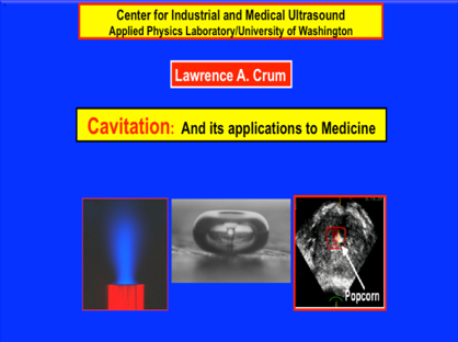

Thursday 13 December 2018
Title: Preparation and characterization of microbubbles et microdroplets by membrane processes for biomedical applications of ultrasounds
Author: Romain MELICH
Summary: The development of various colloidal forms for therapy and diagnosis in medical ultrasound is of great interest since many years. In particular, microbubbles of perfluorocarbon (PFC) are interesting as contrast agents because their gas core is a strong ultrasound reflector. More recently, PFC droplets have been proposed for novel acoustic applications. Indeed, after an acoustic pulse, the ultrasound waves induce a phase change from the liquid state to the gaseous state. This phenomenon is called the acoustic vaporization of droplets. For all these new applications, the development of new techniques of preparation offering a precise control of the particle size is of great necessity. Novel methods of preparation based on membrane devices are particularly interesting for this purpose. The aim of the thesis is the development of new membrane process for the formulation of microbubbles and droplets with tight control of their size, to meet requirements for ultrasound applications in imaging and therapy. In this work, the direct membrane emulsification with a cross-flow membrane module was used for the preparation of microbubbles stabilized by soluble surfactants, while a microkit module was used to fabricate microbubbles stabilized by phospholipids. In a second step, the membrane emulsification by premix was used to formulate monodispersed droplets of PFC. For the various colloidal forms prepared, we documented the influence of the process parameters (pressure, flow rate and shear stress), the formulation parameters (surfactants, type of PFC of the dispersed phase) and the membrane parameters (pore size) on the formation of microbubbles and droplets. Finally, we demonstrated the functionality of microbubbles and droplets, by an acoustic characterization, and showed that they have properties that could be compatible for biomedical applications.
Time: Thursday, December 13th, 2018
Place: Amphithéâtre de physique nucléaire (Paul DIRAC), Campus La Doua
Thursday 6 December 2018
Dear all
The 6th course is now available. It is a technical presentation given by Pr. Chrit MOONEN on «Drug delivery by ultrasound».
It will be followed by a course given by Dr Lawrence A. CRUM entitled «What is cavitation ?». We will then be back in January with more courses.
You can register on Claroline using the following link: clarolineconnect.univ-lyon1.fr.
Hope you’ll enjoy the presentation
We are looking forward to your feedback!
Thank you

Wednesday 28 November 2018
Dear all
The 5th course is now available. It is a technical presentation given Professor Shin-Ichiro UMEMURA on «Ultrasound imaging for high intensity focused ultrasound treatment»
Two more courses will be available before the end of the year. The 6th course will be given by Professor Chrit MOONEN on «Drug delivery by ultrasound», it will then be followed by course 7 given by Dr Lawrence A. CRUM on «What is cavitation ?»
You can register on Claroline using the following link: https://clarolineconnect.univ-lyon1.fr/.
Please note that all the forthcoming courses are indicated on the Agenda.
Hope you’ll enjoy the presentation
We are looking forward to your feedback!
Thank you
Monday 26 November 2018
Author: Stephane Marinesco
Summary: Ce séminaire passera en revue les différents modèles de traumatisme crânien modéré et sévère chez le rat ainsi que les difficultés particulières liées à l’induction d’un trauma sévère chez l’animal. Les mécanismes lésionnels aboutissant à des lésions neurologiques et des déficits comportements seront également discutés.
Time:11:00
Place: Salle de réunion du LabTAU 151 Cours A.Thomas LYON

Wednesday 14 November 2018
Author: Zhishen Sun
Background: Elastography uses shear waves to measure or map the shear modulus for the soft biological tissues [1]. The electromagnetic actuators in magnetic resonance elastography were inexpensive and comparatively simple, and they could work over a wide range of frequency and produce large displacements on the order of 400 μm [2]. However, the electromagnetic actuators currently reported could only work in the MRE but not in ultrasound elastography or optical coherence elastography (OCE) because a strong static magnetic field was needed to create the Lorentz force. On the other hand, induction of shear wave using transcranial magnetic stimulation succeeded in generation of shear wave in soft biological tissues contactlessly [3]. But the shear wave produced by this method was too weak for clinical application - on the order of 0.5 μm. Moreover, this method required additional permanent magnets to create a static magnetic field. This article describes an electromagnetic actuator for the generation of shear waves in the soft media.
Aims: This work aims to confirm the feasibility of generation of shear waves in soft media using an electromagnetic actuator.
Methods: The physics of the force acting on the actuator is explained using classical electromagnetism. The characteristics of the force acting on the actuator are studied by the displacement measurement using an interferometric laser probe (Interferometric probe SH-140, THALES LASER S.A., Orsay, France). The shear wave displacement field generated by the actuator in the PVA phantom is imaged by an ultrasound scanner (Verasonics Vantage 256TM, Washington, USA). The shear wave displacement field was also simulated using the Green function.
Results: The interferometric laser measurement confirms the characteristics of the force acting on the actuator. The actuator generates a shear wave field source of amplitude of 20 μm in the sample of polyvinyl alcohol (PVA) phantom. The shear wave fields created in the PVA phantom by experiments agrees well with that by simulation.
Conclusions: The electromagnetic actuator proves to generate strong shear wave displacement fields.
Time:11:00
Place:Salle de conférence, INSERM 151 Cours Albert Thomas, LYON
Monday 12 November 2018
Dear all
An increasing number of participants has been registering on of the first MOOC on therapeutic ultrasound.
The 4th course is now available. It is a clinical presentation given by Pr Sébastien Crouzet on the use of «HIFU for treating prostate cancer».
In two weeks, it will be followed by the 5th course which will be given by Pr. Shin-Ichiro UMEMURA on «Ultrasound imaging for high intensity focused ultrasound treatment»
You can register on Claroline using the following link: https://clarolineconnect.univ-lyon1.fr/
Hope you’ll enjoy the presentation
Thank you

Thursday 8 November 2018
Elle se tiendra le 8 novembre prochain sur le site Rockefeller (salle Hermann).
La thématique mise en avant cette année portera sur la « Médecine Individualisée », mais comme à l’habitude les doctorants seront encouragés à présenter leurs travaux quel que soit leur thème de recherche, sous forme de communications orales ou de posters. Des enseignants-chercheurs et chercheurs des EPST nouvellement recrutés ou nouvellement arrivés sur le site seront également invités à présenter leurs projets à la communauté, de même que des chercheurs du secteur privé. Tous pourront faire part de leur parcours et de leur expérience lors d’une table-ronde clôturant la journée: l’après-thèse.
Les inscriptions pour cette journée sont ouvertes : https://sfrsle2018.sciencesconf.org

Tuesday 30 October 2018
Thesis defense: Time reversal elastography, mechanical characterization of biological tissues by shear- wave phased array
Date: Tuesday, October 30th, 2018 at 14h
Author: Chadi ZEMZEMI
Location: Conference room, INSERM – Cours Albert Thomas Lyon 3
follow this link for more information

Monday 29 October 2018
Dear all
The third course of the first MOOC on therapeutic ultrasound is now available, it is given by Professor Gail Ter Haar on « Ultrasound induced bio-effects ».
It covers more technical information in order to understand the underlying mechanisms of ultrasound for therapy. It will then be followed by a more clinical case by Professor Sébastien Crouzet on « HIFU for treating prostate cancer ».
You can register on Claroline using the following link: https://clarolineconnect.univ-lyon1.fr/workspaces/tool/properties/21284/subscription/url/generate
Don’t hesitate to head to the forum to discuss with other participants
We are looking forward to your feedback !
Thank you
Friday 19 October 2018
Author: Alexander Doinikov
Time: 14H
Place: AMPHI EST, BÂT HUMANITÉS, INSA DE LYON
Summary: When a body in a fluid is subjected to an acoustic wave field, the scattered wave from the body interacts with the incident wave, giving rise to the co-called acoustic radiation force. This force makes the body migrate in space, and in the case of two or more bodies, causes repulsion or attraction between them. An understanding of this motion is important to applications such as acoustomicrofluidics and medical ultrasonics. The history of theoretical studies of acoustic radiation forces (...)
Thursday 18 October 2018
Author: Thomas Payen
Summary: Harmonic Motion Imaging for pancreatic cancer detection and treatment monitoring- Pancreatic ductal adenocarcinoma (PDA) is one of the deadliest cancers mostly because of late detection due to lack of or unspecific symptoms. One of the main characteristics of PDA is its unusually dense stroma which lower chemotherapy perfusion and efficiency. Developing an imaging technique fit for screening purposes and capable of probing this deep-seeded organ is crucial to improve pancreatic cancer prognosis. During my postdoc at Columbia university, I worked on developing an elastography technique called Harmonic Motion Imaging (HMI) based on an amplitude modulated HIFU signal. The technique was performed in vivo in genetically-engineered mouse models for early tumor detection, pancreatic tissue characterization, and treatment monitoring. HMI was shown to be capable of differentiating healthy pancreas, PDA, and even premalignant pancreatic intraepithelial neoplasias (PanINs), showing that pancreatic tissues get stiffer as the disease progresses. The mechanical response of tumor tissue to HIFU ablation, chemotherapy and stroma depleting enzyme were all successfully monitored with HMI. Finally, HMI was transferred to postsurgical human specimen to demonstrate its feasibility. The technique was capable of detecting both the tumor within perilesional tissue, and regions with normal tissue. In addition, response to neoadjuvant therapy (chemotherapy and radiations) were detected in tumor and normal pancreatic tissue. HMI could potentially prove to be highly beneficial in pancreatic cancer to improve its poor clinical prognosis with early detection, specific characterization, and treatment monitoring
Time:11:00
Place: Salle de réunion LabTAU 151 Cours A.Thomas Lyon
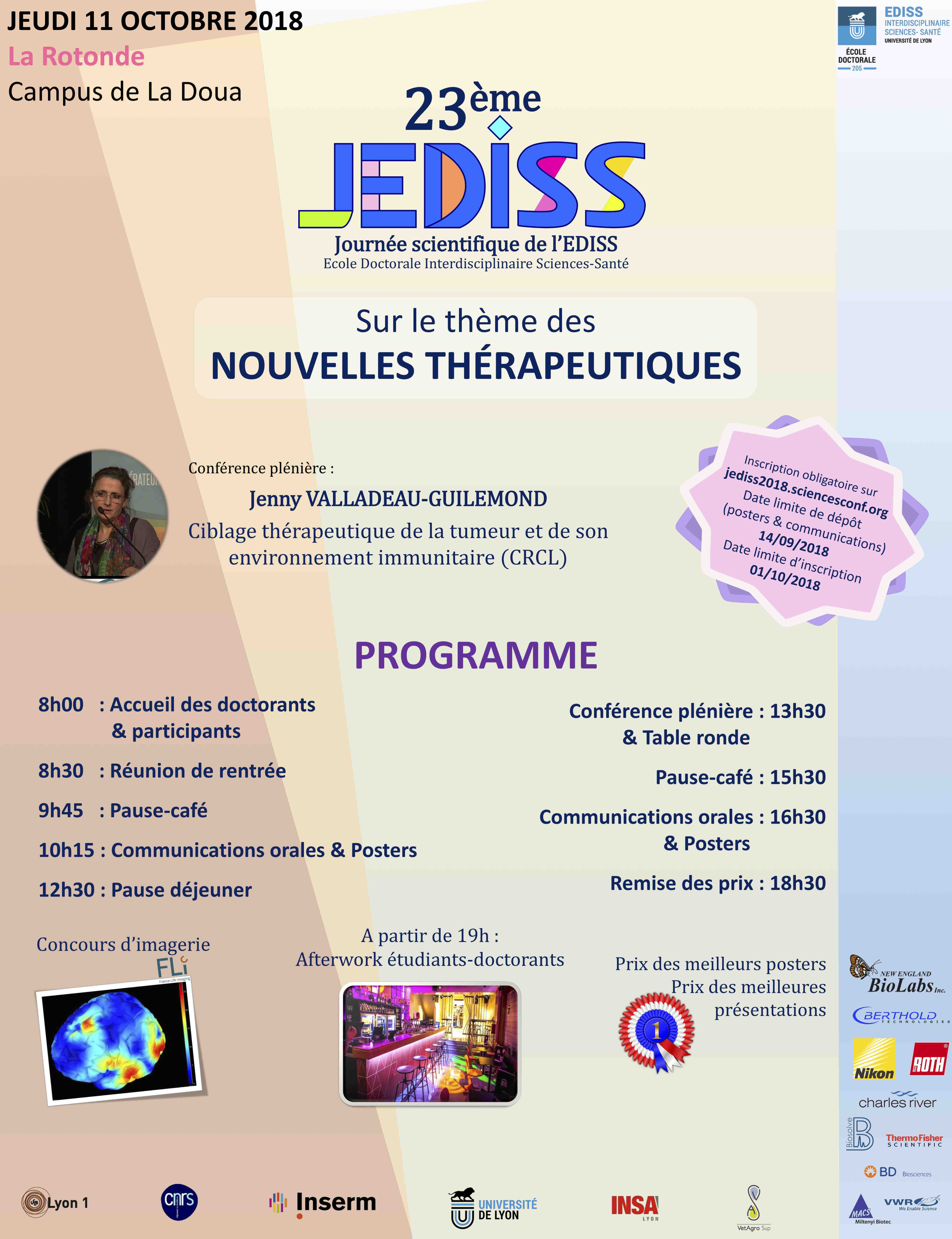
Thursday 11 October 2018
It’s a big pleasure to invite you to for the 23nd scientific day of EDISS, which will take place on Thursday, 11th October at Villeurbanne.
This day will be the occasion to welcome new PhD students and to present them the organization of the doctoral school. It is therefore obligatory for the 1st year PhD students and their supervisors to participate.
To participate, you need to subscribe via https://jediss2018.sciencesconf.org/.
This day is also the occasion for PhD students to present their work via oral communications or posters. We remind you that you have to make a presentation at least once during your PhD.
We hope to see you all!
Monday 1 October 2018
Dear all
The second course of the first MOOC on therapeutic ultrasound is now available
It is given by Oleg Sapozhnikov on « Ultrasound generation and focusing for HIFU therapy ».
You can register on Claroline using the following link:
https://clarolineconnect.univ-lyon1.fr/workspaces/tool/properties/21284/...
We would like to remind that this education program aims at promoting the field of therapeutic ultrasound in its various applications for a targeted audience of Physicians, Students, PhD Students and Patients.
It is open-access and free of charge.
We hope you’ll enjoy it
We are looking forward to your feedback !
Thank you
Thursday 27 September 2018
Les nœuds franciliens, Paris Centre et Paris Sud, du réseau national France Life Imaging, FLI vous invitent à leur journée scientifique le jeudi 27 septembre 2018 à l’Institut de Myologie à Paris.
Cette quatrième édition sera consacrée aux avancées des techniques d'acquisition multimodales en imagerie médicale.
Pour participer, inscrivez-vous jusqu’au 24 septembre inclus à cette adresse : https://fli-pariscentre.sciencesconf.org/
Kahina Abbas pour le comité d'organisation : Olivier Couture, Luc Darrasse, Esther Heinrich, Vincent Lebon, Bernard Van Beers.

Thursday 20 September 2018
Author: Robin Cleveland (Institute of Biomedical Engineering, Department of Engineering Science, University of Oxford)
Summary: Traumatic brain injury has been reportedly widely in the press with respect to athletes subject to head impact and military personnel subject to blast waves. In the case of direct impact there is a typically an acute response, concussion, which can lead to chronic injury to the brain that develops over months and years. The chronic injury manifests itself in issues with memory, mood, and attention. In the case of blast waves there is typically no concussion and yet the chronic brain injury has remarkably similarities. Here we use numerical models and experiments with mice to compare injury induced by blast waves and impact. It is shown that the direct passage of sound energy from the blast wave into the brain tissue does not induce injury or affect memory. Rather it is the oscillation of the head, due to the radiation pressure from the blast wave, that induces shear forces which result in the injury that affects memory.
Time: 11:00
Place: Salle de conference 151 cours albert Thomas 69003 Lyon
Thursday 13 September 2018
Author: Karla P. Mercado-Shekhar, Ph.D.
Summary: Clot stiffness is inversely correlated with rt-PA thrombolytic efficacy in vitro -Predicting thrombolytic susceptibility to recombinant tissue plasminogen activator (rt-PA) a priori could help guide clinical decision-making during acute ischemic stroke treatment and avoid adverse off-target lytic effects. Prior research has shown that the composition and structure of clots impact their mechanical properties and rt-PA thrombolytic efficacy. The goal of the current research was to determine the relationship between clot elasticity and rt-PA thrombolytic efficacy in vitro. Human and porcine mildy and highly retracted clots were fabricated in glass pipettes. The rt-PA thrombolytic efficacy was evaluated in vitro using the percent clot mass loss. A second set of clots were embedded in agar phantoms. Subsequently, the Young’s modulus of each clot was estimated using single-track-location shear wave elasticity imaging. The imaging sequence was implemented using a Verasonics Vantage 256 system equipped with an L7-4 linear array. The results revealed an inverse relationship between the Young’s moduli and clot mass loss, indicating that stiffer clots are more difficult to lyse with rt-PA. This work suggests that clot stiffness may serve as a predictive metric for rt-PA thrombolytic efficacy. Additionally, this presentation will briefly discuss ongoing projects at the Image-guided Ultrasound Therapeutics Laboratories in the University of Cincinnati. These projects include ultrasound-assisted thrombolysis, ultrasound-triggered bioactive gas delivery, and passive cavitation image guidance and monitoring of ultrasound therapeutics.
Time:11:00
Place: Salle de conférence
Wednesday 29 August 2018
Get ready for the release of the first MOOC on therapeutic ultrasound ! On September 2018, the LabTau in collaboration with the Focused Ultrasound Foundation will be launching a MOOC (Massive Online Open Course) dedicated to Therapeutic Ultrasound. In an attempt to promote the field, the targeted audience would first be Medical doctors, students, PhD students and patients. It will be free of charge to all and available online on the educational platform Claroline of the University of Lyon. It is composed of 12 courses (Clinical cases and more technical presentations) given by experts recognized worldwide. Questionnaires and a Forum will enhance interactions between participants. We’ll be happy to update you in September with the latest details for you to access. We hope you’ll join us.
Find more on the following video:
Tuesday 3 July 2018
Author: Corentin CORNU
Summary: Dans l’objectif de limiter les effets destructeurs de l’implosion des bulles de cavitationinertielle, un régime de cavitation purement stable doit potentiellement être visé pour des applications thérapeutiques sensibles comme l’ouverture de la barrière hémato-encéphalique.Cependant, garantir une activité de cavitation stable est difficile puisqu’il peut exister à la fois des bulles oscillantes de manière stable et inertielle au sein d’un même nuage, et qu’une activité de cavitation peut évoluer et basculer d’un état à l’autre au cours du temps,particulièrement en régime pulsé. Il est donc nécessaire pour des applications thérapeutiques de contrôler spatialement et temporellement le phénomène de cavitation en discriminant lesrégimes de cavitation stable ou inertielle. Premièrement, l’étude théorique de la dynamique d’un nuage de bulles de même rayon et uniformément réparties dans l’espace permet de mettre en évidence l’effet de l’interaction entre bulles sur le seuil de cavitation de cavitation stable : il diminue en pression et se décale en fonction de la concentration de bulle. Ainsi, il estmontré qu’il existe une densité de bulle optimale pour l’émission de la composante sous- harmonique. Ensuite, une stratégie de contrôle est développée, basée sur une boucle derétroaction régulant la signature acoustique d’un régime donné de cavitation. L’utilisation de la stratégie d’asservissement permet de discriminer les régimes de cavitation stable et inertielle au cours du temps, mais aussi de garantir une activité de cavitation plus stable temporellement, plus reproductible, et ce pour des énergies acoustiques moyennes délivrées inférieures. Enfin, le processus de contrôle est utilisé expérimentalement pour des applications in-vitro de sonoporation cellulaire. Tout d’abord, une étude de sonoporation en cavitation inertielle régulée met en évidence l’amélioration de la reproductibilité des taux de sonoporation obtenus, et la possibilité de s’affranchir de l’utilisation d’agents de contraste comme agents denucléation. Ensuite, une étude en cavitation stable régulée met en évidence la possibilité de sonoporer des cellules en limitant les activités de cavitation inertielle, et donc potentiellement en limitant la lyse cellulaire.
Time: 14H00
Place: LabTAU (Inserm U1032) Conference Room, 151 Cours Albert Thomas, 69003 Lyon
Tuesday 3 July 2018
Author: Jonathan Mamou
Summary: High-frequency ultrasound (HFU, >20 MHz) and scanning acoustic microscopy (SAM, >200 MHz) offer a means of investigating biological tissue at the microscopic level with spatial resolutions better than 100 μm or 10 μm, respectively. After a brief review of conventional ultrasound imaging, this talk will introduce HFU imaging and present two HFU examples and one SAM example.
Novel HFU imaging methods to form three-dimensional (3D) images of the brain of developing mouse embryos: Many genetically-engineered mice display abnormal development of the central nervous system (CNS) at early embryonic ages and HFU offers great potential for in vivo imaging and characterization. A 34 MHz, five-element annular array was excited using chirp-coded excitation. In utero, in vivo 3D data sets were acquired from more than 100 embryos over five stages, from embryonic days E10.5 to E14.5, and CNS volume renderings were obtained.
The second HFU study focuses on 3D imaging and characterization of lymph nodes freshly-excised from cancer patients. Quantitative-ultrasound (QUS) images were formed and used to detect metastases using a transducer that has a 26-MHz center frequency. Classification results suggest that these QUS methods may provide a clinically-important means of identifying small metastatic foci that might not be detected using standard pathology procedures.
The SAM study focuses on using a novel microscopy system as well as dedicated signal-processing methods to form quantitative images of thin sections of tissue using transducers ranging from 250 MHz to 500 MHz. These techniques yield spatial resolutions better than 8 μm and can be used to assess morphological, acoustical and mechanical tissue properties quantitatively. Data obtained from excised eyes from a guinea pig model of myopia will be presented.
Time:11:00
Place:Salle de réunion duLabTAU u1032 151 Cours A.Thomas , Lyon
Monday 2 July 2018
Une conférence de Mickael TANTER, de l'Institut Langevin (ESPCI Paris), qui sera donnée dans le cadre des conférences du CRNL.
Elle sera accomapgnée par une présentation de Ludovic LECOINTRE, de ICONEUS, une spin-off spécialiste en neuro-imagerie fonctionnelle, notamment par ultrason.
Lundi 2 juillet 2018 11h30
Lieu: Amphitheâtre M Hemingway, Bâtiment IDEE, Pôle Hospitalier Lyon Est Bron
En savoir plus : Mickael_TANTER_Conference_CRNL.pdf
Source : Les conférences du CRNL

Monday 25 June 2018
Author: Oleg Sapozhnikov
Summary: When a focused ultrasound wave of moderate or high intensity in liquid encounters an air interface, a liquid jet emerges from the liquid surface to form what is known as an acoustic fountain. Atomization, or the emission of micro-droplet jets, occurs when the acoustic intensity exceeds a liquid-dependent threshold. It turned out that this effect is related to the recently proposed method of mechanical fractionation of soft biological tissue by HIFU in a process termed boiling histotripsy, wherein the focused ultrasound wave superheats the tissue at the focus, producing a millimeter-size boiling or vapor bubble in several milliseconds. Our hypothesis is that tissue can behave as a liquid such that it forms a fountain and atomization within the vapor bubble produced in boiling histotripsy. High-speed videography was used to observe the formation and atomization of fountains composed of water and other liquids. Similar phenomena were observed in tissues, and, as was shown, they caused erosion of tissue.
Time:11:00
Place:Salle de réunion du LabTAU, INSERM 151 Cours A.Thomas , Lyon
Friday 22 June 2018
Author: Pejman Ghanouni
Summary: Discussion of clinical applications of MR guided focused ultrasound in use or in development at Stanford Hospital
Time:13:30
Place:Salle de réunion du LabTAU, Inserm 151 Cours A.Thomas, Lyon

Thursday 7 June 2018
Author: Simon Bernard
Summary:
Les méthodes d’imagerie basées sur une modélisation numérique complète de la propagation d’ondes dans un milieu hétérogène sont de plus en plus utilisées en sismologie. Ces approches cherchent à identifier la distribution spatiale des paramètres (vitesse de propagation par ex.) du sous-sol en minimisant l’écart entre l’ensemble des sismogrammes enregistrés et les signaux simulés correspondants (inversion de formes d’ondes complètes), ou parfois plus simplement l’écart sur des temps de vol de l’onde entre différentes positions (tomographie de temps de vol).
Dans les deux cas, la précision et la résolution des images sont améliorées par rapport aux méthodes basées sur des approximations (tracé de rayons par ex.) ou sur une simple projection spatiale des signaux (beamforming), au prix d’un coût numérique plus important. Ce coût peut néanmoins être contenu par l’emploi de la méthode dite du champ adjoint, proche du concept de retournement temporel, et n’est plus aujourd’hui une limite aux applications dans le domaine ultrasonore.
Je présenterais deux applications en imagerie médicale : la tomographie ultrasonore des os et l’élastographie par ondes de cisaillement. Dans le premier cas, l’objectif est d’obtenir une carte de la vitesse de propagation des ultrasons dans la section transverse des os long à l’aide d’une antenne circulaire entourant le membre (bras ou jambe), pour obtenir des informations morphologiques et mécaniques sur l’os. Dans la seconde application, il s’agit de corriger les artefacts de diffraction associés à la méthode de reconstruction classique pour obtenir une meilleure évaluation de la rigidité et de la géométrie de lésions dans les tissus mous.
Time:11:00
Place:Salle de Conférence, LabTAU 151 Cours ALbert Thomas, LYON

Thursday 31 May 2018
Afin de promouvoir le rayonnement de son Université et de valoriser les activités de recherche de ses chercheurs(ses), la Métropole de Lyon organise en partenariat avec l’Université de Lyon, le Prix Jeune Chercheur(se) remis à 3 lauréat(e)s distingué(e)s, selon les champs thématiques suivants :
- Thème « biosanté et société», où les enjeux dépassent largement les seuls aspects technologiques, notamment dans les secteurs du cancer et des vaccins, traditionnellement en pointe à Lyon, avec la médecine personnalisée, la croissance de pathologies émergentes, la nécessité de développer des approches avancées de modélisation et simulation.
- Thème « sciences et ingénierie », avec des enjeux comme les industries du futur pour qui les aspects de durabilité et de soutenabilité sont au centre des préoccupations, le partage de l'énergie, les transports.
- Thème « humanités et urbanité », dans une compréhension des métropoles où la technologie, les politiques publiques et les organisations sont tournées vers l'humain. L’analyse des sociétés dans une compréhension de la modernité, du religieux et de la laïcité, de la gestion des croissances économiques et des risques sont étudiés comme des enjeux majeurs.
Date limite de participation : 31 mai 2018.

Thursday 31 May 2018
Author: Pol Grasland-Mongrain
Summary:
Shear waves have been first studied in sismology to probe Earth internal structure. Indeed, a shear wave is a type of mechanical wave with an interesting property: it propagates faster in a harder medium, and vice-versa.
The methods and algorithms developed by sismologists have then been adapted at a much smaller case: the human body. It lead to a biomedical technique, the shear wave elastography. This technique can measure elasticity of organs such as liver, prostate, or breast, through ultrasound imaging or MRI. During my research in the LabTAU in Lyon and in the LBUM in Montreal, I studied a way to induce shear waves using electromagnetic fields, in order to easier measure elasticity of organs in MRI.
Recently, shear wave elastography has also been used to map elasticity at a smaller scale using laser-based imaging methods, on the skin epidermis or the eye cornea for example. In this context, I demonstrated the ability of a laser beam to also induce shear waves at a precise location.
Two years ago, a collaboration between our two labs further decreased the size of the studied sample, at the single cell scale. It needed a fast acquisition, and we used a microscope with an ultrafast camera at 200 000 fps. A proof of concept was done on a mouse oocyte: the cellular sismology was born.
Time:11:00
Place:Salle de réunion du LabTAU 151 Cours Albert Thomas LYON
Friday 25 May 2018
Author: Emilie Franceschini
Summary: Cette présentation porte sur les techniques de spectroscopie ultrasonore permettant d’estimer les structures cellulaires au sein des tissus. Ces techniques permettent de différencier des tissus sains et pathologiques ou de faire le suivi de thérapie. Les tumeurs cancéreuses résultent souvent d'une prolifération de cellules anormales formant une masse qui grossit. Les modèles classiques de diffusion ultrasonore utilisés par les techniques classiques supposent que les diffuseurs sont distribués de façon aléatoire (i.e. des milieux dilués), alors que les tumeurs sont des milieux à fortes concentrations cellulaires. Déterminer les modèles de diffusion adaptés à un amas dense de cellules et identifier les structures cellulaires responsables de la diffusion sont les deux thématiques principales que je développerai. Nous comparons différents modèles de diffusion et discutons de leurs limites à travers des expériences hautes fréquences (10-45 MHz) sur des biofantômes de cellules vivantes ou apoptotiques.
Time:11:00
Place:Salle de réunion, LabTAU u1032 151 Cours A.Thomas, LYON

Wednesday 16 May 2018
Author: Michael Reinwald
Summary: We investigate bone-conducted sound in a short-beaked common dolphin's mandible and attempt to determine whether and to what extent it could contribute to the task of localizing a sound source.
Time:11:00
Place:Salle de conférence, LabTAU, 151 Cours A.Thomas
Tuesday 24 April 2018
Author: Lawrence A. Crum Professor of Bioengineering and Electrical Engineering, Center for Industrial and Medical Ultrasound, University of Washington, Seattle, USA
Dr. Lawrence A. Crum is currently Principal Physicist and Chair of the Department of Acoustics and Electromagnetics in the Applied Physics Laboratory, and Research Professor of Bioengineering and Electrical Engineering at the University of Washington. He has held previous positions at Harvard University, the U. S. Naval Academy and the University of Mississippi, where he was F. A. P. Barnard Distinguished Professor of Physics and Director of the National Center for Physical Acoustics. He has published over 180 articles in professional journals, presented over 200 papers at professional meetings, and holds an honorary doctorate from the Universite Libre de Bruxelles. He is Past President of the Acoustical Society of America and current President of the Board of the International Commission for Acoustics. In June of 2000, he was awarded the Helmholtz-Rayleigh Interdisciplinary Silver Medal of the Acoustical Society of America.
Summary: Acoustic cavitation is a phenomenon that involves the interaction of an acoustic field with a gas bubble in a liquid. Since a bubble has a compressibility many orders of magnitude more than a liquid, the gas bubble can act as a means of concentrating the energy of the acoustic field to a restricted region of time and space. Concentrations of energy on the order of eleven orders of magnitude have been reported. This lecture will introduce cavitation as a physical phenomenon and describe some applications of cavitation to current topics in medical ultrasound.
Time:11:00
Place:Conference room, LabTAU, INSERM 151 Cours Albert Thomas
Monday 26 February 2018
Quelle technologie peut mieux nous faire comprendre l’anatomie d’une cellule, sa fonction ou sa pathologie ? L’élastographie
Un moyen à part entière pour mesurer ultra rapidement les propriétés mécaniques des composantes internes de cellules vivantes : la sismologie cellulaire est née.
C’est la technologie que propose l’équipe de Stefan Catheline, chercheur Inserm au LabTau –laboratoire mixte , Inserm U1032, Université Claude Bernard Lyon 1, Centre Léon Bérard -, en collaboration avec les Hospices Civils de Lyon et l’équipe de Guy Cloutier, chercheur du Centre Hospitalier de Montréal.
Une cellule peut s’assouplir pour accueillir une molécule ou se raidir pour interdire la pénétration d’un corps étranger. Certaines activations électrochimiques induisent même des contractions rapides qui se traduisent par des changements de propriété mécanique de la cellule. Mesurer ces changements instantanés à l’échelle de la cellule permettrait de mieux comprendre la biomécanique cellulaire.
Comment palper une cellule entière instantanément ? Grâce aux ondes élastiques.
Pour élaborer une image d’élasticité cellulaire sous microscope par une approche ondulatoire quantitative, il fallait lever trois verrous technologiques :
- générer des vibrations au moyen d’une micropipette,
- les détecter par une méthode optique,
- enfin en extraire des cartes d’élasticité.
Grâce aux outils mis au point dans le domaine de l’élastographie et de la sismologie, nous mesurons en 1 milliseconde des microséismes sur des ovocytes et en déduisons des cartes d’élasticité.
Dans cet article publié dans PNAS, nous ouvrons donc une nouvelle voie vers le monitoring de la biomécanique cellulaire par microélastographie.
Sources : PNAS MS# 2017-13395RR
Contact chercheur : Stefan Catheline : stefan.catheline@inserm.fr
Contact Presse : Patricia Lefort : patricia.lefort@inserm.fr
Wednesday 31 January 2018
Author: Jeff Elias
William Jeffrey Elias was born in Durham, North Carolina, in 1968. He grew up in Roanoke as the son of a neurologist. He attended Wake Forest University and pursued a Bachelor of Arts degree in chemistry while also playing junior varsity tennis. While in college, he was awarded a scholarship for undergraduate research in chemistry and he graduated Phi Beta Kappa. He attended the University of Virginia for medical school and neurosurgical training. During neurosurgical training, he completed intramural fellowships in neuropathology and spinal surgery before spending a year in Plymouth, England, as a senior registrar. Following residency, he pursued additional training in stereotactic and functional neurosurgery at the Oregon Health Sciences University. In 2002, Dr. Elias returned to the University of Virginia where he is currently the director of stereotactic and functional neurosurgery with a large multidisciplinary program in the surgical treatment of movement disorders, epilepsy and spasticity. He currently sits on the medical advisory board of the International Essential Tremor Foundation (IETF). Dr. Elias’ clinical team includes Caroline Metsch, PAC, Kathy Maynard, RN, and Karen Osteen.
Summary: Therapeutic uses of FUS in the brain.
In the 1950s, neuroscientists and neurosurgeons became interested in using ultrasound to treat disorders deep in the brain. This never gained favor because of the difficulty transmitting acoustic energy through the intact skull. Recent advances in ultrasound transducer technology now enable the precise transmission of ultrasound, through the skull, to deep regions inside the brain. An increasing number of clinical trials now demonstrate the potential efficacy of MRI-guided focused ultrasound for the treatment of movement disorders and other neurologic conditions. This talk will review the current status of focused ultrasound therapy in human brain.
Time:16:00
Place:Conference room

Monday 22 January 2018
Title: Flows generated by a focused ultrasound wave in a liquid medium: Acoustic streaming and solid particles flow.
Summary: The present thesis relates to flows generated by focused ultrasound in homogeneous liquid and in liquid charged with particles. Experimental work in water, using Particle Image Velocimetry (PIV) optical technique and CFD (Computational Fluid Dynamics) numerical modeling using Fluent simulation tool, allow as to study involved hydrodynamic and acoustic phenomena : the fluid acoustic streaming and ultrasound radiation force acting on solid particles. This work is provided in the general context of sono-thrombolysis technique (blood clot destruction by ultrasonic cavitation) optimization, where issues related to thrombolytic agents mixing enhancement and to the risk of clot fragments release in the circulation, arise.
Firstly, this work allowed to evaluate the PIV technique for flow measurement in the particular case of high intensity focused ultrasound field presence, where radiation force is likely to influence the seeding particles movement. Indeed, numerical and experimental results comparison has shown that only 5µm-diameter particles are suitable for streaming measurement. In a second time, we managed to characterize the acoustic streaming in an infinite liquid medium and to compare it with a circular free jet flow. Results related to this characterization showed that the streaming flow, even if it presents structural differences compared to this conventional jet, it has important common characteristics, in particular, related to a self-similarity zone existence. Furthermore, an analysis of radiation force effect on suspended solid particles was carried out. In that, we managed to determine a critical diameter above which solid particles flow is dominated by the ultrasound radiation force rather than the streaming drag force.
Time: 14H30
Place: Salle de conférence du bâtiment INSERM au 151 Cours Albert Thomas, 69003 Lyon
Wednesday 10 January 2018
La médaille de l’innovation a été décernée au Docteur Albert Gelet le 10 janvier 2017 par l’académie de chirurgie validant une activité de recherche fondamentale et clinique qui s’est étalée sur plus de 25 ans concernant le traitement du cancer de la prostate par ultrasons focalisés.
Le traitement du cancer localisé de la prostate par ultrasons focalisés est un traitement mini invasif qui a été développé à partir de 1990 grâce à une collaboration entre 4 partenaires tous basés à Lyon : le service d’urologie et de chirurgie de la transplantation de l’Hôpital Edouard Herriot (Hospices Civils de Lyon), le service de radiologie urinaire de l’Hôpital Edouard Herriot, l’unité INSERM 1032 (application des ultrasons à la thérapie) université Lyon 1, et la société EDAP-TMS. L’objectif était la mise au point d’un traitement aussi peu invasif que possible du cancer localisé de la prostate rendu nécessaire par l’augmentation drastique de son incidence liée à son dépistage par le dosage du PSA (400 000 nouveaux cas en Europe par an dont 70 000 en France) .La plupart des cancers sont maintenant détectés à un stade précoce et un grande nombre de patients présente des cancers dont le risque évolutif à 10 ans (apparition de métastases) est faible ou modéré .Ces patients reçoivent actuellement un traitement radical d’emblée (avec des risques de séquelles urinaires et sexuelles) ou bien sont placés en surveillance active avec traitement différé en cas de progression (avec le risque de réaliser dans certains cas le traitement trop tardivement comme l’a montré l’étude anglaise Protect). L’objectif principal du traitement HIFU est de proposer une alternative à la fois au traitement radical et à le surveillance active à des patients dont la tumeur a été bien évaluée grâce à la combinaison d’une IRM multiparamétrique et de biopsies guidées par l’imagerie IRM.
Wednesday 20 December 2017
Thesis title: Evaluation of a transoesophageal HIFU probe on beating hearts
Summary: Transesophageal HIFU was proposed as an alternative to the cardiac arrhythmias treatments. The work presented in this thesis describes the feasibility of transoeso- phageal thermal ablation of hearts in silico, in vivo and ex vivo, as well as the use of passive elastography as a method of treatment follow-up.
An endoscope integrating a 5MHz 64-element commercial transesophageal echo- cardiography probe and a 8-element HIFU transducer was built. The transducer was cooled at 5°C and ultrasonic beam could be steered over a 15 to 55 mm range.
The first part demonstrates the ability of passive elastoraphy to be used for mo- nitoring thermal injury. The technique has been demonstrated on a simple model of thermosensitive hydrogel, then on biological samples of liver and heart. One study showed the influence of motion on passive elastography.
The prototype was tested on three 30 kg male baboons. Left atrium and ventricles were exposed to repeated continuous sonications (4-15 times during 16s). Passive elastography showed a hardening in the left ventricle after shooting in one of the subjects. T1-weighted and contrast MRI sequences show contradictory results that do not confirm the presence of this lesion.
After a numerical study simulating the influence of motion on the creation of thermal lesions in ultrasound treatment, ultrasound parameters were optimized on a model of isolated beating heart using the Langendorff procedure.
Place: Salle des Thèses (1) de la Faculté de médecine de Rockfeller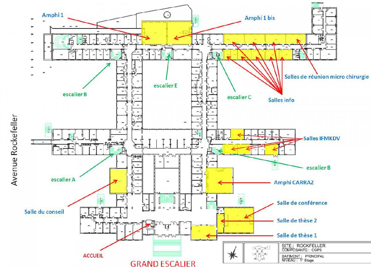
Tuesday 19 December 2017
Thesis title: Ultrasound Microbubbles for Molecular Imaging and Drug Delivery: Detection of Netrin-1 in Breast Cancer & Immunomodulation in Hepatocellular Carcinoma
Summary: Ultrasound has an important role in cancer diagnosis and was further developed for cancer therapy. Diagnostic approaches are required to analyze molecular profiles and enable personalized medicine and therapeutic approaches have to be tumor-specific reducing off-site delivery and side effects. Ultrasound molecular imaging uses microbubbles as ultrasound contrast agents which are functionalized with targeting ligands. Upon intravenous injection, targeted microbubbles bind to molecular markers presented on the tumor endothelium and enable the non-invasive assessment cancer-related biomarkers. In the present thesis, ultrasound molecular imaging was developed for detection of netrin-1, which is upregulated in 70% of metastatic breast cancer and promotes cell survival. A newly developed netrin-1 interference therapy requires the identification of patients who overexpress the target protein and, could benefit from anti-netrin-1 therapy. In vivo imaging of netrin-1 showed a significantly increased imaging signal in netrin-1-positive breast tumors compared to netrin-1-negative breast tumors and normal mammary glands. The results suggest that ultrasound molecular imaging allows accurate detection of netrin-1 on the endothelium of netrin-1-positive tumors and has the potential to become a companion diagnostic for netrin-1 interference therapy in breast cancer patients. Ultrasound-targeted microbubble destruction triggers cavitation and sonoporation thereby permeabilizing the tissue and facilitating local drug delivery. Further, immune cell infiltration and tumor antigen release are induced and trigger anti-tumor immune responses. In the present thesis, ultrasound-targeted microbubble destruction-mediated delivery of anti-cancer microRNA-122 and anti-microRNA-21 is studied for immune response activation in hepatocellular carcinoma, in which the immune microenvironment is deregulated. Tumor lymph nodes showed pro-tumor cytokine downregulation and anti-tumor cytokine upregulation, suggesting an overall positive therapy response with regard to the tumor immunology. The results identified ultrasound-targeted microbubble destruction-mediated miRNA delivery as a potent immuno-modulatory therapeutic approach. In conclusion, ultrasound molecular imaging and ultrasound-targeted microbubble destruction turned out to be powerful techniques that can significantly improve diagnosis and therapy of different types of carcinomas.
Wednesday 13 December 2017
Auteur : Antoine Nordez
Résumé : Les propriétés biomécaniques d’un muscle ou d’un tendon peuvent aisément être évaluées sur modèle animal in vitro ou ex vivo. Ces évaluations sont fondamentales pour analyser la fonction musculaire et comprendre les effets d’entraînements, de désentraînement ou de pathologies. In vivo chez l’homme, des ergomètres peuvent être utilisés, mais ils renseignent sur les propriétés globales d’un système musculo-articulaire, sans différencier les comportements complexes des très nombreux muscles et structures constituant ce système. L’imagerie (principalement l’IRM ou l’échographie) permettent de mesurer des déplacements tissulaires au sein du système mais ne permet pas d’évaluer directement les propriétés mécaniques des muscles, ni les niveaux de tension ou de contrainte qu’ils subissent. En biomécanique musculaire ou tendineuse, nous sommes donc particulièrement intéressés par l’utilisation des méthodes d’élastographie. Lors de cette présentation, une synthèse des travaux réalisés dans notre laboratoire sera réalisée (voir les principales références ci-dessous). Je présenterai en particulier les atouts et limites de la technique commercialisée d’élastographie que nous avons utilisé (Supersonic Shear Imaging). Bien que nous ayons principalement mené des études fondamentales, de nombreuses applications cliniques de l’élastographie peuvent être envisagées sur la base de ces études.
Friday 1 December 2017
Author: Professor Chrit Moonen de « lmage Sciences Institute » at Utrecht.
Summary: The Centre for Image Guided Oncological Interventions (CIGOI) of the UMC Utrecht is an initiative of the Division of Imaging at the University Medical Center, Utrecht, the Netherlands with the central objective to provide real-time image guidance for high precision in the treatment of localized tumors. Within the CIGOI a range of unique image-guided treatment techniques have been developed such as MRI controlled 'High Intensity Focused Ultrasound (MRI guided HIFU), MRI-driven linear accelerator (first prototype installed at UMCU), MRI guided robotic brachytherapy, and intra-arterial radio-embolization with holmium microspheres.
Friday 24 November 2017
Presentation Award to Jennifer Wischhusen for the excellent and outstanding presentation

Wednesday 22 November 2017
Author: Emily White MD Director of operations, Charlottesville, USA
Summary: Emily White, MD joined the Foundation in 2016 as Director of Operations. Prior to joining the Foundation, Dr. White was a private consultant working in operations and business development support in the healthcare and medical start-up space. Her clients have included everything from a 4,000+ employee, publically traded, health care company to a brand new start-up with two employees. Her background includes training in general surgery, leadership positions in several highly technical start-up companies with federal clients, non-profit executive management and over 25 years of grant writing experience. She completed her undergraduate degree in Biology & Anthropology at Smith College, holds certification for Community Conflict Resolution from Loyola Law School, and is a University of Virginia School of Medicine graduate.

Friday 10 November 2017
Author: Professor Paul Barbone, Mechanical Engineering, Boston University.
Summary: Isotropic solid materials can support the propagation of both dilatational waves and distortional waves, the latter better known as shear waves. Propagation of shear waves in soft tissues is a subject of considerable current interest in the biomechanics and biomedical imaging communities. For that purpose, we present an axisymmetric model of elastic shear wave propagation to measure dispersive wave speed and attenuation in homogeneous elastic media. Biomedical imaging applications, however, present much more fun and exotic situations to study shear waves, including in the presence of strong magnetic fields, superposed large poroelastic deformations, strong material property gradients, and medium activation. We use mathematical models to study elastic shear wave propagation in these scenarios.

Wednesday 18 October 2017
Author: Heikki J. Nieminen.
Summary: Non-linear ultrasonics enables actuation of matter from a distance in non-contacting manner. At will this can be achieved with negligible thermal effects. Examples of ultrasonic actuation methods include drug transport, ultrasonic manipulation of nano-fiber drug-delivery systems and acoustic levitation of small animals. The talk will give an insight to such ultrasonic biomedical actuation methods investigated at Medical Ultrasonics Laboratory (MEDUSA), Aalto University, Finland.

![]()