


![]()
Prostate Cancer Imaging
Project Description
About 1 million new cases of prostate cancers are reported each year worldwide. A significant proportion of these cancers are indolent and do not require active treatment. Unfortunately, the evaluation of the aggressiveness of prostate cancer remains difficult. As a consequence, up to 90% of patients may undergo treatment for a cancer unlikely to cause death or even symptoms (Eggener 2015). On the other hand, prostate cancer remains the second cause of cancer-related deaths in men in western countries (Siegel 2015), suggesting that aggressiveness of the disease was underestimated in these patients.
LabTAU develops Quantitative Imaging Computer-Aided Diagnosis to better characterize prostate lesions. Quantitative imaging is the extraction of objective and quantifiable features from medical images. Quantitative imaging provides objective indicators for the assessment of normal or pathologic conditions. It is an essential step into standardizing image interpretation and improving patient care.
This work relies on a longstanding collaboration with the Department of Uro-Radiology at Edouard Herriot Hospital in Lyon, a major center for prostate imaging.
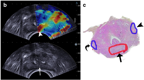
Shear Wave Elastography
Shear Wave Elastography is a quantitative imaging technique for measuring the elasticity (stiffness) of biological tissues, using ultrasound. Our study (Rouvière 2017) demonstrated that Shear Wave Elastography can distinguish prostate malignant and benign tissues.
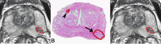
Computer-Aided Diagnosis (CAD)
Multiparametric magnetic resonance imaging (MRI) can help detect prostate cancers, but it suffers from large inter-reader variability and its specificity is poor. In a previous study (Hoang Dinh 2016), we used quantitative multiparametric MR imaging data from patients treated with radical prostatectomy to develop a CAD system for discriminating cancers in the peripheral zone (PZ) with a Gleason score of at least 7. The CAD combined the 10th percentile of the apparent diffusion coefficient (ADC) and the time to peak of enhancement (TTP) at dynamic contrast material–enhanced imaging, and provided good results when cross-validated in data sets from two different manufacturers.
In our most recent study (Hoang Dinh 2018), we assessed the performance of our CAD system in patients referred for multiparametric magnetic resonance (MR) imaging before prostate biopsy. The CAD outperformed the Likert score prospectively assigned by the radiologists.
Staff
- Olivier Rouvière, MD, PhD
- Rémi Souchon, PhD
Selected Publications
- Hoang Dinh et al. Characterization of prostate cancer with Gleason score of at least 7 by using quantitative multiparametric MR imaging: validation of a Computer-Aided Diagnosis system in patients referred for prostate biopsy. Radiology 2018
- Rouvière et al. Stiffness of benign and malignant prostate tissue measured by shear-wave elastography: a preliminary study. Eur Radiol 2017; 27(5):1858-66
- Hoang Dinh et al. Quantitative analysis of prostate multiparametric MR images for detection of aggressive prostate cancer in the peripheral zone: a multiple imager study. Radiology, 2016; 280(1): 117-27
Other references

![]()