


![]()
Streaming and microstreaming
Acoustic streaming and solid particle behavior in focused ultrasound field
Project Description
In sonothrombolysis application, the presence of a fluid medium (blood) subjected to a HIFU field gives rise to acoustic streaming which is flow generation due to acoustic wave propagation in a fluid medium. Acoustic streaming has been shown to be able to improve the mixing in the treatment zone, and therefore to improve the efficiency of thrombolytic agents. This phenomenon is, therefore, to be studied. In this context, acoustic streaming in aquatic medium was investigated. Particle Image Velocimetry (PIV) technique was used to characterize the streaming flow. This technique led us, also, to find that the ultrasonic field, especially the radiation force, can strongly affect solid seeding particle behavior, and consequently, can change the fluid flow nature: Large particles (50µm-diameter) are repelled from the focal zone and gathered at radiation pressure convergence lines on either side of the focus. The calculation of the acoustic radiation pressure applied on these particles explains the observed phenomenon. A simple criterion approximating the diameter threshold, below which seeding particles are not affected by the radiation force and only undergo acoustic streaming drag forces, was then proposed. According to these results and observations, large solid particles are trapped upstream of the focus during ultrasound application. This could be a perspective of studying the radiation force effect on blood clot fragments residence time in the treatment focal zone. In fact, one solution which can make thrombolysis more effective is to trap the fragments, subjected to ultrasound shots, longer in the treatment area in order to completely destroy them and to avoid the risk of secondary embolism.
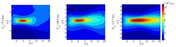
Fig1: Axial velocity field depending on applied acoustic pressure. Focus position: (0,0)
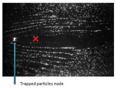
Fig 2: Spatial distribution of 50µm-polyamide seeding particles subjected to focused ultrasound. The red cross indicates the position of the transducer focus
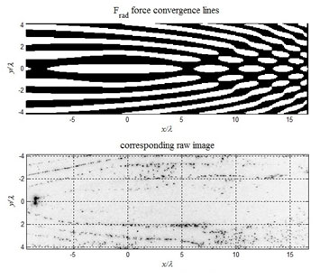
Fig 3: Calculated areas of radiation force convergence (in black) and divergence (in white) VS corresponding inversed raw image (particles in black)
Staff
LabTau Staff
-
Rafika Ben Haj Slama (PhD student)
-
Bruno Gilles (Associate Professor)
-
Jean-Christophe Bera (Professor)
Collaborations
-
Maher Ben Chiekh (Associate Professor, LESTE, Monastir, Tunisia)
Publications

![]()